ANAT2341 Lab 6 - Fetal: Difference between revisions
(Created page with "{{ANAT2341Lab6}} ==Fetal Growth== {| | <Flowplayer height="320" width="285" autoplay="true">fetal growth.flv</Flowplayer><br> Quicktime...") |
mNo edit summary |
||
| (3 intermediate revisions by the same user not shown) | |||
| Line 1: | Line 1: | ||
{{ANAT2341Lab6}} | {{ANAT2341Lab6}} | ||
==Fetal Growth== | ==Fetal Growth== | ||
{| | {| | ||
| | | <html5media height="480" width="400">File:fetal growth.mp4</html5media> | ||
< | |||
[[ | :'''Links:''' [[Fetal Development Movie]] | [[Media:fetal growth.mp4|MP4 version]] | [[Fetal Development]] | ||
| | | | ||
In this cartoon movie of fetal growth, observe the changing relative sizes of the head and | In this cartoon movie of fetal growth, observe the changing relative sizes of the head, body and limbs. | ||
| Line 15: | Line 17: | ||
Finally consider fetal changes that are occurring in auditory pathway. | Finally consider fetal changes that are occurring in auditory pathway. | ||
|} | |} | ||
==Fetal Head Growth== | |||
[[File:Fetal_head_growth_circumference_graph01.jpg|600px]] | |||
'''Head Circumference Growth''' (both gestational and post-fertilisation ages are shown) | |||
==Skull Ossification== | ==Skull Ossification== | ||
These are 2 views of the same 12 week 92 mm CRL human fetus head, double stained to show both cartilage (blue) and newly-formed bone (red). The head undergoes two different forms of ossification (endochondral and intramembranous) in separate regions of the skull. | These are 2 views of the same 12 week 92 mm CRL human fetus head, double stained to show both cartilage (blue) and newly-formed bone (red). The head undergoes two different forms of ossification (endochondral and intramembranous) in separate regions of the skull. | ||
[[File:Fetal head lateral.jpg|400px]] | {| | ||
|-bgcolor="CEDFF2" | |||
! Lateral view (external) | |||
! Medial view (internal) | |||
|- | |||
| [[File:Fetal head lateral.jpg|400px]] | |||
| [[File:Fetal head medial.jpg|400px]] | |||
|- | |||
| Note the distribution of new bone formation by '''intramembranous ossification''' in the plates of the cranial vault, temporal bone, orbit, upper jaw (maxilla) and lower jaw (mandible) regions. Bony regions in the lower jaw (mandible) region also show spaces where tooth formation is occurring. | |||
| Note the distribution of cartilage from the nasal region through the base of the skull showing '''endochondral ossification''', also occuring in the atlas/axis (with new bone forming). See also the original Meckel's cartilage within the newly forming bony mandible. | |||
|} | |||
[[File:Skull_superior.gif]] | |||
[[File: | Late Fetal Skull | ||
===Mandible Ossification=== | |||
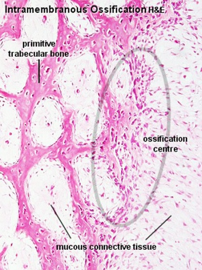
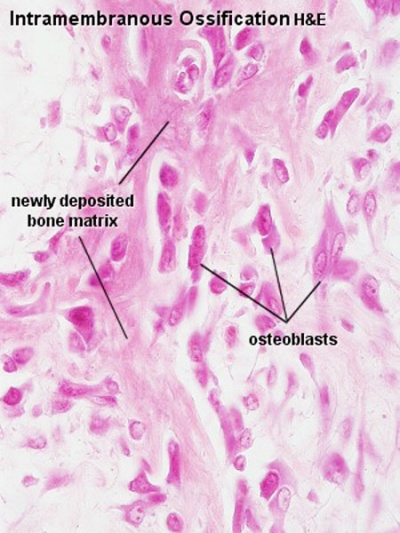
[[File:Ossification_centre.jpg|400px]] [[File:Intramembranous_ossification_centre.jpg|400px]] | |||
==Face== | ==Face== | ||
The cartilage template of the mandible and the base of the skull are replaced by early bone development. | The cartilage template of the mandible and the base of the skull are replaced by early bone development. | ||
[[File:Fetal_head_section.jpg|400px]] | [[File:Fetal_head_section.jpg|400px]] [[File:Fetal_head_section_01.jpg|400px]] | ||
Selected midline medial head view showing key features of head musculoskeletal and neurological development. | Selected midline medial head view showing key features of head musculoskeletal and neurological development. | ||
| Line 45: | Line 62: | ||
==Palate Development== | ==Palate Development== | ||
[[File:Bailey140.jpg|thumb|Early Fetal Palate]] | |||
Secondary palate formation is the growth of the palatal shelves towards the midline. | |||
{| border='0px' | {| border='0px' | ||
|-bgcolor="CEDFF2" | |||
! Inferior view | |||
! Anterior view | |||
|- | |- | ||
| | | width=360px|<html5media height="400" width="400">File:Palate_001.mp4</html5media> | ||
| width=360px|<html5media height="400" width="400">File:Palate_002.mp4</html5media> | |||
|- | |- | ||
| [[File:Palate_001_icon.jpg]] | |||
| [[File:Palate_002_icon.jpg]] | |||
|} | |||
[[File:Bailey140.jpg|thumb|Early Fetal Palate]] | |||
{| border='0px' | |||
| valign="bottom"|{{Palate 1}} | |||
| valign="bottom"|{{Palate 2}} | |||
| '''Palate Overview''' | |||
Embryonic | |||
* '''week 4''' - pharyngeal arch formation, first pharngeal arch contributes mandible and maxilla. | |||
* '''week 6 - 7''' - primary palate formation maxillary processes and frontonasal prominence. | |||
Fetal | |||
* '''week 9''' - secondary palate shelves fuse, separating oral and nasal cavities. | |||
|} | |} | ||
Week 10 Gestational Age ({{GA}} week 12) | |||
{| | |||
| [[File:Fetal week 10 hard palate 07.jpg|400px]] | |||
hard palate | |||
| [[File:Fetal_week_10_soft_palate_03.jpg|400px]] | |||
soft palate | |||
|} | |||
{{ANAT2341Lab6}} | {{ANAT2341Lab6}} | ||
Latest revision as of 12:50, 9 September 2016
| Lab 6: Introduction | Trilaminar Embryo | Early Embryo | Late Embryo | Fetal | Postnatal | Abnormalities | Online Assessment |
Fetal Growth
<html5media height="480" width="400">File:fetal growth.mp4</html5media>
|
In this cartoon movie of fetal growth, observe the changing relative sizes of the head, body and limbs.
|
Fetal Head Growth
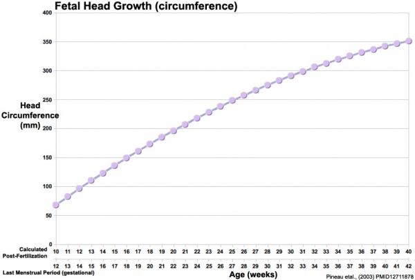
Head Circumference Growth (both gestational and post-fertilisation ages are shown)
Skull Ossification
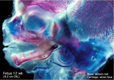
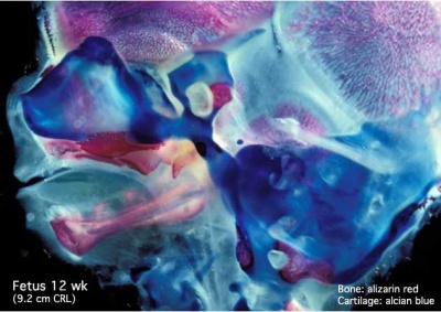
These are 2 views of the same 12 week 92 mm CRL human fetus head, double stained to show both cartilage (blue) and newly-formed bone (red). The head undergoes two different forms of ossification (endochondral and intramembranous) in separate regions of the skull.
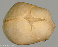
Late Fetal Skull
Mandible Ossification
Face
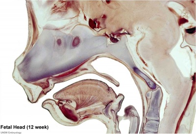
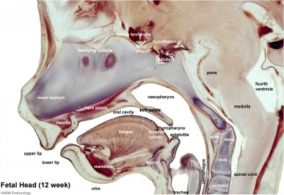
The cartilage template of the mandible and the base of the skull are replaced by early bone development.
Selected midline medial head view showing key features of head musculoskeletal and neurological development.
Note extensive nasal cartilage, nasal conchae, pituitary, secondary palate, oral cavity, tongue, mandible, hyoid, choana, oropharynx.
Also note the developing tongue musculature and its mandibular attachment site.
Note that the cranial vault, the portion of the skull enclosing the brain, ossifies by a unique bone formation process, intramembranous ossification.
Because the head contains many different structures also review notes on Special Senses (eye, ear, nose), Respiration (pharynx), Integumentary (Teeth), Endocrine (thyroid, parathyroid, pituitary) and Musculoskeletal (tongue, skull).
Palate Development
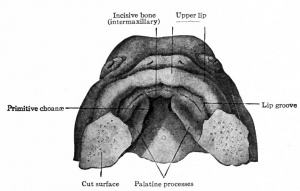
Secondary palate formation is the growth of the palatal shelves towards the midline.
| Inferior view | Anterior view |
|---|---|
| <html5media height="400" width="400">File:Palate_001.mp4</html5media> | <html5media height="400" width="400">File:Palate_002.mp4</html5media> |
|
|
Palate Overview
Embryonic
Fetal
|
Week 10 Gestational Age (GA week 12)
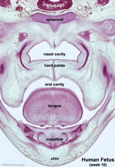
hard palate |
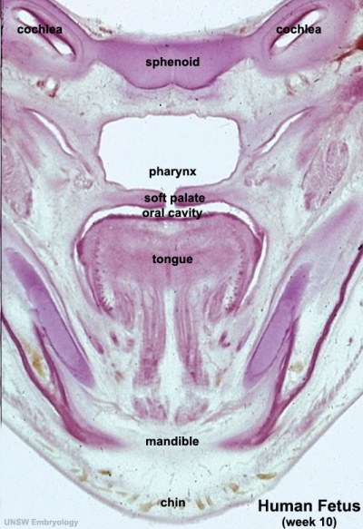
soft palate |
| Lab 6: Introduction | Trilaminar Embryo | Early Embryo | Late Embryo | Fetal | Postnatal | Abnormalities | Online Assessment |
Glossary Links
- Glossary: A | B | C | D | E | F | G | H | I | J | K | L | M | N | O | P | Q | R | S | T | U | V | W | X | Y | Z | Numbers | Symbols | Term Link
Cite this page: Hill, M.A. (2024, April 19) Embryology ANAT2341 Lab 6 - Fetal. Retrieved from https://embryology.med.unsw.edu.au/embryology/index.php/ANAT2341_Lab_6_-_Fetal
- © Dr Mark Hill 2024, UNSW Embryology ISBN: 978 0 7334 2609 4 - UNSW CRICOS Provider Code No. 00098G
