ANAT2341 Lab 4 - Implantation and Villi Development: Difference between revisions
No edit summary |
No edit summary |
||
| Line 32: | Line 32: | ||
File:Gray0027.gif | File:Gray0027.gif | ||
</gallery> | </gallery> | ||
==Villi Development== | |||
==Chorionic Villi== | |||
{| | |||
|- | |||
| '''Primary villi''' | |||
'''Week 2''' - first stage of chorionic villi development, trophoblastic shell cells (syncitiotrophoblasts and cytotrophoblasts) form finger-like extensions into maternal decidua. | |||
| [[File:Gray0036.gif]] | |||
|- | |||
| '''Secondary villi''' | |||
'''Week 3''' - second stage of chorionic villi development, extraembryonic mesoderm grows into villi, covers entire surface of chorionic sac. | |||
Basal region will form chorionic plate. | |||
| [[File:Gray0037.gif]] | |||
|- | |||
| '''Tertiary villi''' | |||
'''Week 4''' - third stage of chorionic villi development, mesenchyme differentiates into blood vessels and cells, forms arteriocapillary network, fuse with placental vessels, developing in connecting stalk. | |||
| [[File:Gray0031.jpg|300px]] | |||
|} | |||
* '''stem villi''' - or anchoring villi, cytotrophoblast cells attached to maternal tissue. | |||
* '''branched villi''' - grow from sides of stem villi, region of main exchange, surrounded by maternal blood in intervillous spaces. | |||
* '''terminal villi''' - not active outgrowths caused by proliferation of the trophoblast. Passive protrusions induced by capillary coiling due to growth of the fetal capillaries within the mature intermediate villi (third trimester). | |||
* '''chorionic plate''' - region of membrane at the base of the villi through which placental arteries and vein passes. | |||
==First trimester and term chorionic villi== | |||
===Anchoring Villi=== | |||
[[File:Placenta_anchoring_villi.jpg]] | |||
===Floating Villi=== | |||
[[File:Human_placental_villi_cartoon_01.jpg|800px]] | |||
<gallery> | |||
File:Placenta anchoring villi.jpg|Placenta anchoring villi | |||
File:Placental villi.jpg|Villi first trimester | |||
File:Placental villi 5.jpg|Villi at term | |||
File:Placental_villi_3.jpg|Villi at term | |||
</gallery> | |||
==Virtual Slide== | |||
We will now look at an example of first trimester placentation in a Virtual Slide. | |||
[http://vslides.unsw.edu.au/VirtualSlideV2.nsf/id/74A731 Virtual Slides - Placenta] | |||
Please note that there are additional slides listed in the current set, only the first placenta slide and the cord cross-section will be covered in detail in the practical class. | |||
==Chorionoic Villi Location== | |||
Originally villi cover entire chorionic surface and then become restricted to decidua basalis region forming 2 regions: | |||
# '''Frondosum''' - "leafy" where villi are mainly located. | |||
# '''Capsularis''' - smooth chorion, where villi are absent or not abundant. | |||
{| | |||
| [[File:Embryo-membranes_stage_11.jpg|300px]] | |||
| [[File:Stage18_bf10.jpg|500px]] | |||
|- | |||
| <center>Week 4 (Stage 11)</center> | |||
| <center>Week 7 (Stage 18)</center> | |||
|} | |||
==Additional Information== | |||
[[File:Placenta_and_explant_model.jpg|thumb|600px|Placental villi cartoon]] | |||
===Cytotrophoblast Layer=== | |||
There is a new interpretation of the changes that are occuring in the cytotrophoblast (CTB) layer during early to full-term human placenta development. Traditionally the interpretation was that the cytotrophoblast layer thinned and became discontinuous towards term. The thinning is thought due to the epithelium surface expanding at a faster rate than its volume. Two recent studies suggest that while the cytotrophoblast layer does indeed thin, it does not become discontinuous. | |||
===Syncytiotrophoblast Layer=== | |||
The syncytiotrophoblast (STB) layer forms the epithelial covering of the entire villous tree. These cells are multinucleated, terminally-differentiated syncytium formed by the fusion of the underlying progenitor cytotrophoblast (CTB) cells. The process is described as "syncytialization" and is mediated by syncytin-1, an envelope protein of a human endogenous retrovirus W (HERV-W). The differentiation is regulated by chorionic gonadotropin (hCG) and the fusion of cytotrophoblast cells is ongoing during placental development. | |||
Cellular parts derived from the syncytiotrophoblasts (apoptotic nuclei and microparticulate debris) can be shed into the maternal blood in which they are bathed. The apototic process appears to be part of the fusion mechanism between cytotrophoblast and the overlying multinucleate syncytiotrophoblast layer. | |||
Studies have suggested that these cells are transcriptionally inactive. A recent study using a number of different detection techniques now suggests that at least some of the cells nuclei may still be transcriptionally inactive. | |||
===Mesenchymal Villi=== | |||
Mesenchymal villi generate all other villous types: | |||
* immature intermediate villi | |||
* stem villi | |||
* mature intermediate villi | |||
* terminal villi | |||
Mesenchymal villi continuously form out of the trophoblastic sprouts throughout pregnancy and have been considered the basis for growth and differentiation of the villous trees. | |||
===Human Villi Timeline=== | |||
This is far more than even for additional information section [[Talk:BGDA_Practical_Placenta_-_Villi_Development|Timeline Table]] | |||
<gallery> | |||
File:Gray0034.jpg|Decidua and villi location | |||
File:Bailey495.jpg|Chorionic villi | |||
</gallery> | |||
{{ANAT2341Lab4}} | {{ANAT2341Lab4}} | ||
{{2012ANAT2341}} | {{2012ANAT2341}} | ||
Revision as of 23:14, 14 August 2012
| ANAT2341 Lab 4: Introduction | Implantation and Villi | Decidua and Cord | Abnormal Placenta | Cardiovascular | Online Assessment | Group Project |
Implantation

Human Embryo Day 8 to 9
| Implantation | Chorionic Cavity | Week 3 | Amniotic Cavity |
| Quicktime | Flash | Quicktime | Flash | Quicktime | Flash | Quicktime | Flash |
Week 3 embryo


Villi Development
Chorionic Villi
| Primary villi
Week 2 - first stage of chorionic villi development, trophoblastic shell cells (syncitiotrophoblasts and cytotrophoblasts) form finger-like extensions into maternal decidua. |

|
| Secondary villi
Week 3 - second stage of chorionic villi development, extraembryonic mesoderm grows into villi, covers entire surface of chorionic sac. Basal region will form chorionic plate. |

|
| Tertiary villi
Week 4 - third stage of chorionic villi development, mesenchyme differentiates into blood vessels and cells, forms arteriocapillary network, fuse with placental vessels, developing in connecting stalk. |

|
- stem villi - or anchoring villi, cytotrophoblast cells attached to maternal tissue.
- branched villi - grow from sides of stem villi, region of main exchange, surrounded by maternal blood in intervillous spaces.
- terminal villi - not active outgrowths caused by proliferation of the trophoblast. Passive protrusions induced by capillary coiling due to growth of the fetal capillaries within the mature intermediate villi (third trimester).
- chorionic plate - region of membrane at the base of the villi through which placental arteries and vein passes.
First trimester and term chorionic villi
Anchoring Villi

Floating Villi


Placenta anchoring villi
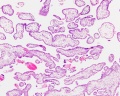
Villi first trimester
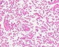
Villi at term
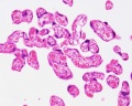
Villi at term


Virtual Slide
We will now look at an example of first trimester placentation in a Virtual Slide.
Please note that there are additional slides listed in the current set, only the first placenta slide and the cord cross-section will be covered in detail in the practical class.
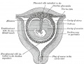
Chorionoic Villi Location
Originally villi cover entire chorionic surface and then become restricted to decidua basalis region forming 2 regions:
- Frondosum - "leafy" where villi are mainly located.
- Capsularis - smooth chorion, where villi are absent or not abundant.

|

|
Additional Information

Cytotrophoblast Layer
There is a new interpretation of the changes that are occuring in the cytotrophoblast (CTB) layer during early to full-term human placenta development. Traditionally the interpretation was that the cytotrophoblast layer thinned and became discontinuous towards term. The thinning is thought due to the epithelium surface expanding at a faster rate than its volume. Two recent studies suggest that while the cytotrophoblast layer does indeed thin, it does not become discontinuous.
Syncytiotrophoblast Layer
The syncytiotrophoblast (STB) layer forms the epithelial covering of the entire villous tree. These cells are multinucleated, terminally-differentiated syncytium formed by the fusion of the underlying progenitor cytotrophoblast (CTB) cells. The process is described as "syncytialization" and is mediated by syncytin-1, an envelope protein of a human endogenous retrovirus W (HERV-W). The differentiation is regulated by chorionic gonadotropin (hCG) and the fusion of cytotrophoblast cells is ongoing during placental development.
Cellular parts derived from the syncytiotrophoblasts (apoptotic nuclei and microparticulate debris) can be shed into the maternal blood in which they are bathed. The apototic process appears to be part of the fusion mechanism between cytotrophoblast and the overlying multinucleate syncytiotrophoblast layer.
Studies have suggested that these cells are transcriptionally inactive. A recent study using a number of different detection techniques now suggests that at least some of the cells nuclei may still be transcriptionally inactive.
Mesenchymal Villi
Mesenchymal villi generate all other villous types:
- immature intermediate villi
- stem villi
- mature intermediate villi
- terminal villi
Mesenchymal villi continuously form out of the trophoblastic sprouts throughout pregnancy and have been considered the basis for growth and differentiation of the villous trees.
Human Villi Timeline
This is far more than even for additional information section Timeline Table
Decidua and villi location
Chorionic villi


| ANAT2341 Lab 4: Introduction | Implantation and Villi | Decidua and Cord | Abnormal Placenta | Cardiovascular | Online Assessment | Group Project |
- 2012 Course: Week 1 Lecture 1 Lecture 2 Lab 1 | Week 2 Lecture 3 Lecture 4 Lab 2 | Week 3 Lecture 5 Lecture 6 Lab 3 | Week 4 Lecture 7 Lecture 8 Lab 4 | Week 5 Lecture 9 Lecture 10 Lab 5 | Week 6 Lecture 11 Lecture 12 Lab 6 | Week 7 Lecture 13 Lecture 14 | Lab 7 | Week 8 Lecture 15 Lecture 16 Lab 8 | Week 9 Lecture 17 Lecture 18 Lab 9 | Week 10 Lecture 19 Lecture 20 Lab 10 | Week 11 Lecture 21 Lecture 22 Lab 11 | Week 12 Lecture 23 Lecture 24 Lab 12
Glossary Links
- Glossary: A | B | C | D | E | F | G | H | I | J | K | L | M | N | O | P | Q | R | S | T | U | V | W | X | Y | Z | Numbers | Symbols | Term Link
Cite this page: Hill, M.A. (2024, April 19) Embryology ANAT2341 Lab 4 - Implantation and Villi Development. Retrieved from https://embryology.med.unsw.edu.au/embryology/index.php/ANAT2341_Lab_4_-_Implantation_and_Villi_Development
- © Dr Mark Hill 2024, UNSW Embryology ISBN: 978 0 7334 2609 4 - UNSW CRICOS Provider Code No. 00098G