Cardiovascular Movies
These simple cartoons show, almost in developmental sequence, aspects of cardiac development. Each linked page is supported by simple descriptions of events shown in the movie (Note - Quicktime and Flash links are 2 versions of the same movie, the Flash movies will not play on iPads or iPhones).
|
|
Whole Embryo Folding
The animation shows how the folding of the whole embryo in week 3-4 (GA week 5-6) brings the cardiac region into its anatomical location.
|
|
|
Heart Looping
The early heart tube buckles and folds as it grows in length, forming the initial shape of the future heart.
|
|
|
Ventricular Growth
As the ventricular region grows there is a realignment of the ventricles in the developing heart tube.
|
|
|
Atrial Septation
The division of the atria occurs by the growth of two separate overlapping septa in the embryonic period. Note that both septa are not complete and allow blood to pass between the two atria during the entire prenatal period (embryonic and fetal).
|
|
|
Outflow Septation
The ventricles initially separate by growth of the inferior muscular wall. Later the superior region separates by outgrowth of membranous regions that extend into the outflow tract. This third and final septation, leads to a separation of the pulmonary and aortic arch circulations. (Note that a shunt, the ductus arteriosus will persist through the entire prenatal period (embryonic and fetal).
|
The historic movies below include an audio commentary of cardiac development events (note the audio is unfortunately quite poor).
Cardiac Embryology Tutorial
Next work through the online Cardiac Embryology Tutorial.
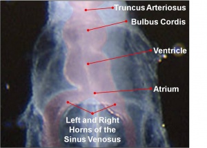
Human embryo stage 10 heart tube
This website is an educational resource designed to teach human cardiac embryology and is a Medicine ILP project carried out by Phoebe Norville. Heart development represents an important area of both embryological and clinical studies, predominantly due to the high incidence of congenital heart disease in the community. Therefore this website aims to teach cardiac embryology to students of all ages with varying degrees of knowledge in the area. The module contains three different levels:
- Basic - begin here if you are new to heart embryology (high school level)
- Intermediate - begin here if you have some background in heart embryology (university level)
- Advanced - begin here after you have completed the earlier levels and have a good background in heart embryology (university level)
Your initial knowledge level determines your start level. Each level is then divided into a sequence of units/pages that roughly correspond to the sequence of heart development. The more detailed, the more pages; basic level has only 3 units, intermediate has 7 units, and the advanced module has 9 units. You can work through each level in sequence or jump to a more detailed level within each unit.
Navigation
Each unit also has two sets of navigation panels and a timeline, to help work through the cardiac embryology modules.
| Top navigation panel
This panel allows you to easily move between pages within the same level or to jump to the beginning of a different level; green is the basic module, yellow the intermediate, and red the advanced.
|
|
| Bottom navigation panel
This panel allows you to easily move through the information sequentially as well as moving to the same content in a different level of complexity.
|
|
| Timelines
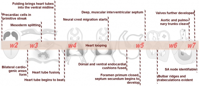
A timeline will appear at the top of each page to help you understand the order and context of the events occurring in cardiac embryology.
|
|
Many of the concepts taught in embryology are best represented with animations or diagrams. The animations in this module are played using Adobe Flash Player. Most of the pictures appear in the units as thumbnails. To see the images more clearly simply click on the picture which will take you to the original-sized version. To return to the module simply click the back button in your browser window.
Basic module
Intermediate module
Advanced module
Glossary Links
- Glossary: A | B | C | D | E | F | G | H | I | J | K | L | M | N | O | P | Q | R | S | T | U | V | W | X | Y | Z | Numbers | Symbols | Term Link
Cite this page: Hill, M.A. (2024, April 16) Embryology ANAT2341 Lab 11 - Heart. Retrieved from https://embryology.med.unsw.edu.au/embryology/index.php/ANAT2341_Lab_11_-_Heart
- What Links Here?
- © Dr Mark Hill 2024, UNSW Embryology ISBN: 978 0 7334 2609 4 - UNSW CRICOS Provider Code No. 00098G


