ANAT2341 Lab 10 - Online Assessment 2015: Difference between revisions
| Line 60: | Line 60: | ||
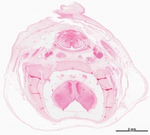
The lens of the eye is derived from surface ectoderm. Said ectoderm forms a lens/optic placode in the head region which then invaginates to form a lens pit and then later a lens vessel. Lens fibres then develop and are surrounded by a lens capsule. The main function of the lens is to focus light onto the retina. | The lens of the eye is derived from surface ectoderm. Said ectoderm forms a lens/optic placode in the head region which then invaginates to form a lens pit and then later a lens vessel. Lens fibres then develop and are surrounded by a lens capsule. The main function of the lens is to focus light onto the retina. | ||
'''Embryology link''' [[Vision- Lens Development]] --Development Overview | '''Embryology link''' [[Vision - Lens Development]] --Development Overview | ||
Revision as of 13:20, 17 October 2015
| Embryology - 23 Apr 2024 |
|---|
| Google Translate - select your language from the list shown below (this will open a new external page) |
|
العربية | català | 中文 | 中國傳統的 | français | Deutsche | עִברִית | हिंदी | bahasa Indonesia | italiano | 日本語 | 한국어 | မြန်မာ | Pilipino | Polskie | português | ਪੰਜਾਬੀ ਦੇ | Română | русский | Español | Swahili | Svensk | ไทย | Türkçe | اردو | ייִדיש | Tiếng Việt These external translations are automated and may not be accurate. (More? About Translations) |
Individual Assessment
- Place your work on this page under a sub-sub-heading of your ROI.
- Add your own sub-sub-heading below any existing student ROI.
| About this Assessment | ||||||||||||||||||||||||||||||||||||
|---|---|---|---|---|---|---|---|---|---|---|---|---|---|---|---|---|---|---|---|---|---|---|---|---|---|---|---|---|---|---|---|---|---|---|---|---|
| A demonstration of this assessment will be given in the practical class. Below in the collapsible table are examples of links from a virtual slide. There is also a permalink help page.
| ||||||||||||||||||||||||||||||||||||


Student ROIs
This is a sub-sub-heading
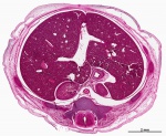
Cochlear Duct
link to permalink image:| Cochlear Duct
The cochlear duct is an fluid filled cavity inside the cochlea. It located between the tympanic duct and the vestibular duct, and between the basilr membrane and reissner's memebrane. It derived from otic placode, otic vesicle, and originated from surface ectoderm.
embryology link Sensory - Hearing and Balance Development --Inner Ear
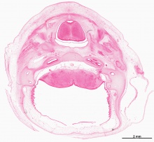
Semicircular Canal
link to permalink image:| Semicircular Canal
The semicircular canals are part of the inner ear.They are lined with cilia and filled with endolymph which is a liquid substance. Every time the head moves, the endolymph moves the cilia and this movements of the cilia are communicated to the brain. As a result, the brain knows how to keep the body balanced, regardless of the posture.
embryology link [Semicircular Canal-http://www.ncbi.nlm.nih.gov/pubmedhealth/PMHT0024846/] -- Inner Ear
Lens of the Eye
Link to permalink image: Anterior portion of the Lens of the embryonic eye
The lens of the eye is derived from surface ectoderm. Said ectoderm forms a lens/optic placode in the head region which then invaginates to form a lens pit and then later a lens vessel. Lens fibres then develop and are surrounded by a lens capsule. The main function of the lens is to focus light onto the retina.
Embryology link Vision - Lens Development --Development Overview
| ANAT2341 Lab 10: Introduction | Early Embryo | Late Embryo | Fetal | Postnatal | Abnormalities | Online Assessment |
- 2015 Course: Week 2 Lecture 1 Lecture 2 Lab 1 | Week 3 Lecture 3 Lecture 4 Lab 2 | Week 4 Lecture 5 Lecture 6 Lab 3 | Week 5 Lecture 7 Lecture 8 Lab 4 | Week 6 Lecture 9 Lecture 10 Lab 5 | Week 7 Lecture 11 Lecture 12 Lab 6 | Week 8 Lecture 13 Lecture 14 Lab 7 | Week 9 Lecture 15 Lecture 16 Lab 8 | Week 10 Lecture 17 Lecture 18 Lab 9 | Week 11 Lecture 19 Lecture 20 Lab 10 | Week 12 Lecture 21 Lecture 22 Lab 11 | Week 13 Lecture 23 Lecture 24 Lab 12 | 2015 Projects: Three Person Embryos | Ovarian Hyper-stimulation Syndrome | Polycystic Ovarian Syndrome | Male Infertility | Oncofertility | Preimplantation Genetic Diagnosis | Students | Student Designed Quiz Questions | Moodle page