ANAT2241 Endocrine System: Difference between revisions
No edit summary |
|||
| Line 15: | Line 15: | ||
File:Thyroid_histology_002.jpg|Thyroid (high power) | File:Thyroid_histology_002.jpg|Thyroid (high power) | ||
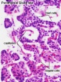
File:Parathyroid_histology_001.jpg|Parathyroid (low power) | File:Parathyroid_histology_001.jpg|Parathyroid (low power) | ||
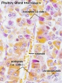
File:Parathyroid_histology_002.jpg|Parathyroid (high power) | File:Parathyroid_histology_002.jpg|Parathyroid (high power)File:Pancreatic islet.png|Pancreatic islet | ||
</gallery> | |||
===Pituitary=== | |||
<gallery> | |||
File:Pituitary histology 001.jpg|Pituitary - adenohypophysis | File:Pituitary histology 001.jpg|Pituitary - adenohypophysis | ||
File:Pituitary histology 002.jpg|Pituitary - adenohypophysis | File:Pituitary histology 002.jpg|Pituitary - adenohypophysis | ||
File:Pituitary histology 003.jpg|Pituitary - neurohypophysis | File:Pituitary histology 003.jpg|Pituitary - neurohypophysis | ||
</gallery> | |||
===Adrenal=== | |||
<gallery> | |||
File:Adrenal histology 001.jpg|Adrenal - Cortex and Medulla | File:Adrenal histology 001.jpg|Adrenal - Cortex and Medulla | ||
File:Adrenal histology 002.jpg|Adrenal - Cortical Zones | File:Adrenal histology 002.jpg|Adrenal - Cortical Zones | ||
File:Adrenal histology 003.jpg|Adrenal - Zona Reticularis and Medulla | File:Adrenal histology 003.jpg|Adrenal - Zona Reticularis and Medulla | ||
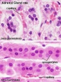
Adrenal histology 004.jpg | |||
Adrenal histology 005.jpg | |||
Adrenal histology 006.jpg | |||
Adrenal histology 007.jpg | |||
Adrenal histology 008.jpg | |||
Adrenal histology 009.jpg | |||
Adrenal histology 010.jpg | |||
Adrenal histology 011.jpg | |||
</gallery> | </gallery> | ||
{{Pituitary Histology}} | {{Pituitary Histology}} | ||
Revision as of 15:21, 15 February 2013

| ANAT2241 This practical support page content is not part of the virtual science practical class and provides additional information for student self-directed learning purposes. All practical class pages are located on Moodle - ANAT2241 |
Aims
Currently a template page - this notice removed when complete.
Links: Virtual Slides - Endocrine System | Endocrine support page
Histology
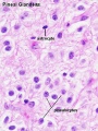
Pineal (high power)
Thyroid (low power)
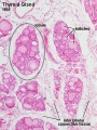
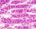
Thyroid (high power)
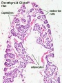
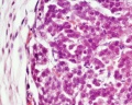
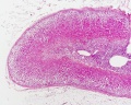
Parathyroid (low power)
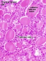
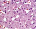
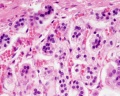
Pancreatic islet


Pituitary
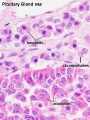
Pituitary - adenohypophysis
Pituitary - adenohypophysis
Pituitary - neurohypophysis


Adrenal
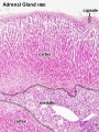
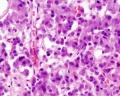
Adrenal - Cortex and Medulla
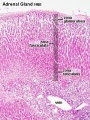
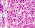
Adrenal - Cortical Zones
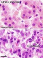
Adrenal - Zona Reticularis and Medulla


- Pituitary Histology: Pituitary overview | Anterior H&E | Anterior H&E | Anterior labeled | PAS/O Overview | Acidophils | Basophils | Posterior labeled | Posterior unlabeled | Histology Stains | BGD - Endocrine Histology | Pituitary Development
- Thyroid Links: low power image | high power image | unlabeled human image | unlabeled sheep image | thyroid
Course Links
- Histology Glossary: A | B | C | D | E | F | G | H | I | J | K | L | M | N | O | P | Q | R | S | T | U | V | W | X | Y | Z | ANAT2241 Support | Histology | Histology Stains | Embryology Glossary
| Common Histology Stains | ||||||||||||||||||||||||||||||||||||||||||||||||||||||||||||||||||||||||||||||||||||||||||||||||||||||||||||||||||||||||||||||||||||||||||||||||
|---|---|---|---|---|---|---|---|---|---|---|---|---|---|---|---|---|---|---|---|---|---|---|---|---|---|---|---|---|---|---|---|---|---|---|---|---|---|---|---|---|---|---|---|---|---|---|---|---|---|---|---|---|---|---|---|---|---|---|---|---|---|---|---|---|---|---|---|---|---|---|---|---|---|---|---|---|---|---|---|---|---|---|---|---|---|---|---|---|---|---|---|---|---|---|---|---|---|---|---|---|---|---|---|---|---|---|---|---|---|---|---|---|---|---|---|---|---|---|---|---|---|---|---|---|---|---|---|---|---|---|---|---|---|---|---|---|---|---|---|---|---|---|---|---|
| ||||||||||||||||||||||||||||||||||||||||||||||||||||||||||||||||||||||||||||||||||||||||||||||||||||||||||||||||||||||||||||||||||||||||||||||||
| ||||||||||||||||||||||||||||||||||||||||||||||||||||||||||||||||||||||||||||||||||||||||||||||||||||||||||||||||||||||||||||||||||||||||||||||||
Practical Support
- Pages can be accessed from any internet connected computer.
ANAT2241 Support Links: The Virtual Microscope | Covering and Lining Epithelia | Glandular Epithelia | CT Components | CT Types | Bone, Bone Formation and Joints | Muscle | Nervous | Blood | Eye | Cardiovascular | Respiratory | Integumentary | Gastrointestinal | Gastrointestinal Organs | Lymphatic and Immune | Endocrine | Urinary | Female Reproductive | Male Reproductive | Histology Stains | Histology Drawings | Practicals Health and Safety 2013 | Moodle - 2019
ANAT2241 This practical support page content is not part of the science practical class and provides only background information for student self-directed learning purposes.
Cite this page: Hill, M.A. (2024, April 24) Embryology ANAT2241 Endocrine System. Retrieved from https://embryology.med.unsw.edu.au/embryology/index.php/ANAT2241_Endocrine_System
- © Dr Mark Hill 2024, UNSW Embryology ISBN: 978 0 7334 2609 4 - UNSW CRICOS Provider Code No. 00098G