ANAT2241 Covering and Lining Epithelia: Difference between revisions
mNo edit summary |
|||
| (31 intermediate revisions by the same user not shown) | |||
| Line 1: | Line 1: | ||
{{ANAT2241 header}} | {{ANAT2241 header}} | ||
==General Objective== | |||
[[File:Histology_terminology_cartoon.jpg|thumb]] | |||
To learn the structure and function of covering and lining epithelia. | |||
Associated lecture slides: [[Media:ANAT2241 Epithelia Lecture2018.pdf|Covering and Lining Epithelia (17.51 MB PDF)]] | [[Media:ANAT2241 Epithelia Lecture2018 4 slides.pdf|4 slides/page]] | |||
===Specific Objectives=== | ===Specific Objectives=== | ||
| Line 16: | Line 17: | ||
To identify, draw and label the various types of epithelium (simple, stratified and pseudostratified) and to discuss some of the special functional features associated with their structure. | To identify, draw and label the various types of epithelium (simple, stratified and pseudostratified) and to discuss some of the special functional features associated with their structure. | ||
Virtual Slides: [https://moodle.telt.unsw.edu.au/mod/book/view.php?id=789982&chapterid=100720 Covering and Lining Epithelia] | |||
{| | {| | ||
| Line 25: | Line 30: | ||
| Epithelia sectioning appearance | | Epithelia sectioning appearance | ||
|} | |} | ||
==Histology== | ==Histology== | ||
| Line 57: | Line 61: | ||
===Stratified epithelium=== | ===Stratified epithelium=== | ||
Oesophagus stratified squamous non-keratinized epithelium. | |||
<gallery> | <gallery> | ||
File:Oesophagus histology 03.jpg | File:Oesophagus histology 03.jpg | ||
| Line 63: | Line 69: | ||
File:Oesophagus histology 02.jpg | File:Oesophagus histology 02.jpg | ||
</gallery> | </gallery> | ||
===Transitional epithelium=== | ===Transitional epithelium=== | ||
| Line 72: | Line 76: | ||
Urinary bladder transitional epithelium | Urinary bladder transitional epithelium | ||
| | | [[File:Ureter_histology_001.jpg|400px]] | ||
Ureter transitional epithelium | |||
|} | |||
'''Unlabeled Images''' | |||
<gallery> | <gallery> | ||
File:Bladder histology 001.jpg | File:Bladder histology 001.jpg|Low power image showing underlying muscle layers. | ||
File:Bladder histology 002.jpg | File:Bladder histology 002.jpg| | ||
File:Bladder histology 003.jpg | File:Bladder histology 003.jpg| | ||
File:Bladder histology 004.jpg | File:Bladder histology 004.jpg| | ||
</gallery> | </gallery> | ||
==Electron Micrographs== | |||
===Epithelia Specialisations - Cilia=== | |||
{| | |||
|+ '''Respiratory Epithelium - Cilia''' | |||
|- | |||
| valign=bottom|{{SlideRespiratoryEpitheliumEM01}} | |||
| valign=bottom|{{SlideRespiratoryEpitheliumEM02}} | |||
|- | |||
| '''Scanning Electron micrograph''' | |||
<br> | |||
[[File:RespiratoryEpitheliumSEM01.jpg|300px]] | |||
This respiratory epithelium surface view by scanning electron microscope (SEM) shows the 3 dimensional structure of the cilia protruding into the airspace. | |||
| | |||
<br>These cilia move the overlying mucus that has trapped particles, bacteria and viruses, entering the respiratory tract as shown in the movie below.{{#pmid:18250199|PMID18250199}} | |||
<html5media height="350" width="800">File:Mouse trachea 01.mp4</html5media> | |||
|} | |} | ||
[[File:Cilium cartoon.jpg]] | |||
===Epithelia Specialisations - Villi=== | |||
[[File:Gastrointestinal villi and crypts cartoon.jpg|300px]] | |||
===Simple Squamous Epithelium=== | |||
{| | |||
|+ '''Endothelium - Simple Squamous''' | |||
|- | |||
| [[File:Blood capillary EM 02.jpg|600px]] | |||
| An electron micrograph of a capillary blood vessel with white blood cell located in the lumen. | |||
The endothelium is an example of a '''simple squamous epithelium'''. | |||
Note the nuclei of the endothelial cells bulging into the vessel lumen. | |||
* The white blood cell nucleus shape suggests that it is a [[:File:Monocyte_01.jpg|Monocyte]]. | |||
Scale bar 1 μm {{Osmium}} | |||
|} | |||
===Epithelia Specialisations - Junctions=== | |||
{| | |||
|+ '''Epithelial Junctions''' | |||
|- | |||
| [[File:Epithelial junctions EM01.jpg|600px]] | |||
| Junctional complex between two cells in the epithelium of the intestinal mucosa ({{rat}}), intestinal lumen is towards the top of image. | |||
These 2 epithelial cells have a range of different junctional complexes along their lateral contact membranes. Half (hemi-) versions of these junctions also occur on the cell basal membrane where it attaches to the extracellular matrix (basal lamina, visible in EM), not shown in this image. | |||
* '''Tight junction''' - (zonula occludens), located nearest the lumen, extends from arrow 1 to arrow 2. The narrowing of the apparent intercellular "gap" (~90 A) is clearly visible, but the fusion line of the two apposed membranes cannot be clearly distin- guished at this magnification. Note that there is relatively little accumulation of dense cytoplasmic material along this part of the complex. | |||
* '''Adherens junction''' - (zonula adhaerens) intermediate junction extends from arrow 2 to arrow 3. A relatively wide intercellular space (~200 A) is maintained throughout the junction. Extensive condensation of cytoplasmic fibrils occurs as a fine feltwork along either side of the junction. This condensation is continuous with the terminal web (tw) into which the filamentous rootlets (r) of the microvilli penetrate. Plate-like densifications within the cytoplasmic feltwork can be seen along part of the junction, especially along the right side (pi). | |||
* '''Desmosome''' - marked by arrows 4 and 5. This element is characterized by a wide intercellular space (~240 A) bisected by an intermediate line ('''id'''). Bundles of cytoplasmic fibrils ('''fd'''), coarser (diameter ~80 A) and more distinct than those of the terminal web, converge into dense plates ('''pd''') on each side of the desmo- some. These plates are separated from the inner leaflets of the cell membrane by a zone of low density. Similar fibrils ('''if''') appear throughout the remainder of the field below the terminal web. | |||
Between the intermediate junction and the desmosome, the two apposed cell membranes are separated by an irregular space of varying width and show membrane invaginations and associated vesicles ('''v'''). The trilaminar structure of the cell membrane ('''cm''') shows clearly along the microvilli ('''my'''), (wherever the membrane is sectioned normally), and within the desmosome. It can also be made out, though less regularly, along the lateral cell margins (e.g., at unnumbered arrow). | |||
Note that the luminal membrane is nearly symmetrical, the outer leaflet being only slightly thinner and less dense than the inner leaflet, whereas the lateral membrane is definitely asymmetrical. The total thickness of all three layers is about 110 A along the apical surface of the cell but measures only about 70 to 80 A along the lateral intercellular spaces. Note also the fluffy dense material ('''fro''') (probably mucus) associated with the tips and sides of the microvilli. | |||
Specimen fixed in 2 per cent {{Osmium}} in acetate-Veronal buffer (pH 7.6) ; and embedded in Epon. Pb(OH)2-stained section. X 96,000. {{Osmium}} | |||
|} | |||
{| | |||
|+ '''Epithelial Junction - Desmosome''' | |||
|- | |||
| File:Desmosome 02.jpg|600px]] | |||
|} | |||
{{ANAT2241 footer}} | {{ANAT2241 footer}} | ||
Latest revision as of 12:37, 9 April 2019

| ANAT2241 This practical support page content is not part of the virtual science practical class and provides additional information for student self-directed learning purposes. All practical class pages are located on Moodle - ANAT2241 |
General Objective

To learn the structure and function of covering and lining epithelia.
Associated lecture slides: Covering and Lining Epithelia (17.51 MB PDF) | 4 slides/page
Specific Objectives
- To identify covering and lining epithelia.
- To understand the terms endothelium, and mesothelium.
- To classify epithelia on the basis of cell morphology and cell arrangement (e.g. layering), noting the general absence of blood vessels in epithelia.
- To study the surface specializations of some epithelial cells: brush or striated border (microvilli), non-motile stereocilia and motile cilia and to recognise these specialisation in electron micrographs.
Learning Activities
To identify, draw and label the various types of epithelium (simple, stratified and pseudostratified) and to discuss some of the special functional features associated with their structure.
Virtual Slides: Covering and Lining Epithelia

|

|
| Epithelia cell shape | Epithelia sectioning appearance |
Histology
Epithelium forms continuous layers of cells that cover surfaces and line cavities of the body.
Simple epithelium

Venule with endothelium, a simple squamous epithelium lining. |

Thyroid follicles, with a simple cuboidal epithelium surrounding each follicle. |
Pseudostratified epithelium

Trachea lined with a pseudostratified epithelium. |

Testis ductus epididymidis lined with a pseudostratified epithelium. |

Testis ductus epididymidis lined with a pseudostratified epithelium.
Stratified epithelium
Oesophagus stratified squamous non-keratinized epithelium.


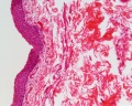
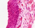
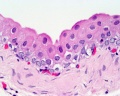
Transitional epithelium

Urinary bladder transitional epithelium |

Ureter transitional epithelium |
Unlabeled Images
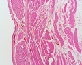
Low power image showing underlying muscle layers.


Electron Micrographs
Epithelia Specialisations - Cilia
|
| ||||||
| Scanning Electron micrograph
This respiratory epithelium surface view by scanning electron microscope (SEM) shows the 3 dimensional structure of the cilia protruding into the airspace. |
<html5media height="350" width="800">File:Mouse trachea 01.mp4</html5media> |


Epithelia Specialisations - Villi

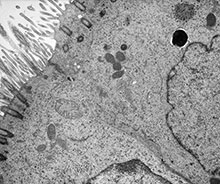
Simple Squamous Epithelium

|
An electron micrograph of a capillary blood vessel with white blood cell located in the lumen.
The endothelium is an example of a simple squamous epithelium. Note the nuclei of the endothelial cells bulging into the vessel lumen.
Scale bar 1 μm (Stain - Osmium) |
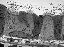
Epithelia Specialisations - Junctions

|
Junctional complex between two cells in the epithelium of the intestinal mucosa (rat), intestinal lumen is towards the top of image.
These 2 epithelial cells have a range of different junctional complexes along their lateral contact membranes. Half (hemi-) versions of these junctions also occur on the cell basal membrane where it attaches to the extracellular matrix (basal lamina, visible in EM), not shown in this image.
Note that the luminal membrane is nearly symmetrical, the outer leaflet being only slightly thinner and less dense than the inner leaflet, whereas the lateral membrane is definitely asymmetrical. The total thickness of all three layers is about 110 A along the apical surface of the cell but measures only about 70 to 80 A along the lateral intercellular spaces. Note also the fluffy dense material (fro) (probably mucus) associated with the tips and sides of the microvilli.
|
| 600px]] |
Course Links
- Histology Glossary: A | B | C | D | E | F | G | H | I | J | K | L | M | N | O | P | Q | R | S | T | U | V | W | X | Y | Z | ANAT2241 Support | Histology | Histology Stains | Embryology Glossary
| Common Histology Stains | ||||||||||||||||||||||||||||||||||||||||||||||||||||||||||||||||||||||||||||||||||||||||||||||||||||||||||||||||||||||||||||||||||||||||||||||||
|---|---|---|---|---|---|---|---|---|---|---|---|---|---|---|---|---|---|---|---|---|---|---|---|---|---|---|---|---|---|---|---|---|---|---|---|---|---|---|---|---|---|---|---|---|---|---|---|---|---|---|---|---|---|---|---|---|---|---|---|---|---|---|---|---|---|---|---|---|---|---|---|---|---|---|---|---|---|---|---|---|---|---|---|---|---|---|---|---|---|---|---|---|---|---|---|---|---|---|---|---|---|---|---|---|---|---|---|---|---|---|---|---|---|---|---|---|---|---|---|---|---|---|---|---|---|---|---|---|---|---|---|---|---|---|---|---|---|---|---|---|---|---|---|---|
| ||||||||||||||||||||||||||||||||||||||||||||||||||||||||||||||||||||||||||||||||||||||||||||||||||||||||||||||||||||||||||||||||||||||||||||||||
| ||||||||||||||||||||||||||||||||||||||||||||||||||||||||||||||||||||||||||||||||||||||||||||||||||||||||||||||||||||||||||||||||||||||||||||||||
Practical Support
- Pages can be accessed from any internet connected computer.
ANAT2241 Support Links: The Virtual Microscope | Covering and Lining Epithelia | Glandular Epithelia | CT Components | CT Types | Bone, Bone Formation and Joints | Muscle | Nervous | Blood | Eye | Cardiovascular | Respiratory | Integumentary | Gastrointestinal | Gastrointestinal Organs | Lymphatic and Immune | Endocrine | Urinary | Female Reproductive | Male Reproductive | Histology Stains | Histology Drawings | Practicals Health and Safety 2013 | Moodle - 2019
ANAT2241 This practical support page content is not part of the science practical class and provides only background information for student self-directed learning purposes.
Cite this page: Hill, M.A. (2024, April 23) Embryology ANAT2241 Covering and Lining Epithelia. Retrieved from https://embryology.med.unsw.edu.au/embryology/index.php/ANAT2241_Covering_and_Lining_Epithelia
- © Dr Mark Hill 2024, UNSW Embryology ISBN: 978 0 7334 2609 4 - UNSW CRICOS Provider Code No. 00098G