ANAT2241 Blood

| ANAT2241 This practical support page content is not part of the virtual science practical class and provides additional information for student self-directed learning purposes. All practical class pages are located on Moodle - ANAT2241 |
Introduction

The circulating blood is a liquid connective tissue consisting of cells (red and white blood cells), fragments of cells (platelets) and liquid (plasma). The different cell types are all derived from haemopoietic stem cells located in the bone marrow. Red blood cells (RBCs) have a metabolic role, in carrying oxygen to tissues and carbon dioxide to the lungs. White blood cells (WBCs or leukocytes) have a role in the body’s defence, and are an important clinical indicator of disease.
Virtual Slide Box: 1. Human Blood Smear Slide
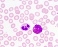
Find an area in the smear where the red blood cells are spread out and individual cells can be identified.
Identify:
- Red blood cells (7-8 um diameter anucleate biconcave disc)
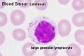
- White blood cells: neutrophils, eosinophils, basophils, lymphocytes and monocytes (basophils are normally rare).
Note the presence or absence of granules, shape of the nucleus and relative cell sizes. Also identify platelets.
Virtual Slide Box: 2. Bone Marrow Smear Slide
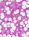
- Do not attempt to identify all the cells in the bone marrow smear, but compare its appearance with that of the blood smear.
- Haematopoiesis (hematopoiesis) is the process of blood cell differentiation and occurs mainly in the bone marrow.
- This bone marrow smear will contain a large number of differentiating blood cells: band cells and normoblasts.
- The largest cells visible are megakaryocytes, which are responsible for platelet production.
Lymphocyte differentiation begins in the bone marrow and continues in central lymphoid organs (bone marrow - B cells and thymus - T cells), then in the peripheral lymphoid organs (lymph nodes, spleen).
Blood Histology
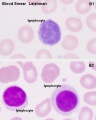
Lymphocyte
Lymphocyte
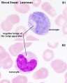
Monocyte
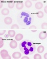
Neutrophil
Neutrophil and Eosinophil
Bone Marrow


Course Links
- Histology Glossary: A | B | C | D | E | F | G | H | I | J | K | L | M | N | O | P | Q | R | S | T | U | V | W | X | Y | Z | ANAT2241 Support | Histology | Histology Stains | Embryology Glossary
| Common Histology Stains | ||||||||||||||||||||||||||||||||||||||||||||||||||||||||||||||||||||||||||||||||||||||||||||||||||||||||||||||||||||||||||||||||||||||||||||||||
|---|---|---|---|---|---|---|---|---|---|---|---|---|---|---|---|---|---|---|---|---|---|---|---|---|---|---|---|---|---|---|---|---|---|---|---|---|---|---|---|---|---|---|---|---|---|---|---|---|---|---|---|---|---|---|---|---|---|---|---|---|---|---|---|---|---|---|---|---|---|---|---|---|---|---|---|---|---|---|---|---|---|---|---|---|---|---|---|---|---|---|---|---|---|---|---|---|---|---|---|---|---|---|---|---|---|---|---|---|---|---|---|---|---|---|---|---|---|---|---|---|---|---|---|---|---|---|---|---|---|---|---|---|---|---|---|---|---|---|---|---|---|---|---|---|
| ||||||||||||||||||||||||||||||||||||||||||||||||||||||||||||||||||||||||||||||||||||||||||||||||||||||||||||||||||||||||||||||||||||||||||||||||
| ||||||||||||||||||||||||||||||||||||||||||||||||||||||||||||||||||||||||||||||||||||||||||||||||||||||||||||||||||||||||||||||||||||||||||||||||
Practical Support
- Pages can be accessed from any internet connected computer.
ANAT2241 Support Links: The Virtual Microscope | Covering and Lining Epithelia | Glandular Epithelia | CT Components | CT Types | Bone, Bone Formation and Joints | Muscle | Nervous | Blood | Eye | Cardiovascular | Respiratory | Integumentary | Gastrointestinal | Gastrointestinal Organs | Lymphatic and Immune | Endocrine | Urinary | Female Reproductive | Male Reproductive | Histology Stains | Histology Drawings | Practicals Health and Safety 2013 | Moodle - 2019
ANAT2241 This practical support page content is not part of the science practical class and provides only background information for student self-directed learning purposes.
Cite this page: Hill, M.A. (2024, April 19) Embryology ANAT2241 Blood. Retrieved from https://embryology.med.unsw.edu.au/embryology/index.php/ANAT2241_Blood
- © Dr Mark Hill 2024, UNSW Embryology ISBN: 978 0 7334 2609 4 - UNSW CRICOS Provider Code No. 00098G