AE Practical - Neural Histology: Difference between revisions
(→Aims) |
|||
| Line 23: | Line 23: | ||
==Key concepts== | ==Key concepts== | ||
[[File:Neuron cartoon.jpg|thumb|'''A Neuron''' - the functional unit of thenervous system]] | |||
The brain and spinal cord comprise the central nervous system (CNS). The nerves that emerge from the spinal cord and brain to pass to parts of the body are the peripheral nervous tissue (PNS). Nervous tissue, with many interconnections, forms a complex system of neuronal communication within the body and is specialized for detecting stimuli, integrating functions, controlling effectors and higher functions. Nervous tissue consists of cell bodies, cell processes (nerves), and neuroglia (supporting cells). | The brain and spinal cord comprise the central nervous system (CNS). The nerves that emerge from the spinal cord and brain to pass to parts of the body are the peripheral nervous tissue (PNS). Nervous tissue, with many interconnections, forms a complex system of neuronal communication within the body and is specialized for detecting stimuli, integrating functions, controlling effectors and higher functions. Nervous tissue consists of cell bodies, cell processes (nerves), and neuroglia (supporting cells). | ||
Revision as of 10:27, 24 September 2012
Introduction

- Draft Page
 This page provides histology support information for central nervous system structure. Disclaimers
|

|
Aims
- Obtain an understanding of the normal histological appearance of selected central and peripheral nervous system tissues namely spinal cord, cerebellum and peripheral nerve.
- To examine unique microscopic characteristics of each of the nervous tissues.
- To introduce the histology and neuropathology associated with cerebral infarction and haemorrhage.
Key concepts

The brain and spinal cord comprise the central nervous system (CNS). The nerves that emerge from the spinal cord and brain to pass to parts of the body are the peripheral nervous tissue (PNS). Nervous tissue, with many interconnections, forms a complex system of neuronal communication within the body and is specialized for detecting stimuli, integrating functions, controlling effectors and higher functions. Nervous tissue consists of cell bodies, cell processes (nerves), and neuroglia (supporting cells).
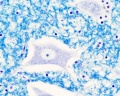
Neurons: Structural and Functional units of the nervous system
These cells (around 12 billion) are responsible for the receptive, integrative, and motor functions of the nervous system. They can generate nerve impulses (irritability), and can transmit these impulses along their processes (conductivity). They range in diameter from 5 to 150 μm and contain 3 parts: a cell body, multiple dendrites and a single axon.
- Cell body (soma, perikaryon) is the region of the neuron containing a large pale-staining spherical, nucleus with a conspicuous nucleolus and perinuclear cytoplasm.
- Dendrites project from the cell body and are specialized for receiving (afferent) stimuli from sensory cells, axons and other neurons which are then transmitted towards the soma.
- Axons arise as a single thin process extending longer distances from the cell body than the dendrite. As with dendrites, the terminals of the axon are branching and terminate in end bulbs (terminal boutons), which come close to another cell and form a synapse.
Practical class activities

Brain Histology
Overview Cortex (mouse)
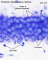
Cortex (mouse)
- Brain histology 03.jpg
- Brain histology 04.jpg


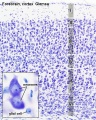
Developing Brain Histology

- Human embryo developing cortex, Week 8, Carnegie stage 22.
- Inset (upper right) shows section overview and approximate level of section (red line).
- Thin layer outer called cortical plate will eventually form the adult brain cortex.
- Other underlying layers are part of the development process and will continue to supply cells to the cortex through fetal period, these layers will eventually be almost completely lost.
- Developing cerebrum layer thicknesses are shown in microns.
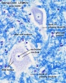
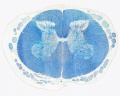
Spinal Cord Histology
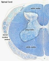
Overview
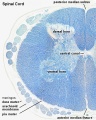
Overview
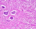
Grey matter
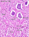
Grey matter (HE)
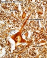
Grey matter (silver)
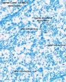
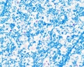
White matter
Overview unlabeled
Grey matter unlabeled 1
Grey matter unlabeled 2
White matter unlabeled 1
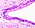
Ependymal cells unlabeled


- Spinal Cord: Overview 1 | Overview 2 | Overview animation | Grey matter | Grey matter | Grey matter | White matter | Overview unlabeled | Grey matter unlabeled 1 | Grey matter unlabeled 2 | White matter unlabeled 1 | Ependymal cells unlabeled
Terms
- artifact - changes and distortions introduced to the normal tissue structure by the histological processing. Common artifacts include: folds (gives the tissue a darker appearance), tears (rips in the tissue can be seen in epithelia), shrinkage when tissues loose mainly liquid through histological processing, and cuts often used in tissue preparation.
Glossary Links
- Glossary: A | B | C | D | E | F | G | H | I | J | K | L | M | N | O | P | Q | R | S | T | U | V | W | X | Y | Z | Numbers | Symbols | Term Link
Cite this page: Hill, M.A. (2024, April 20) Embryology AE Practical - Neural Histology. Retrieved from https://embryology.med.unsw.edu.au/embryology/index.php/AE_Practical_-_Neural_Histology
- © Dr Mark Hill 2024, UNSW Embryology ISBN: 978 0 7334 2609 4 - UNSW CRICOS Provider Code No. 00098G