AE Practical - Neural Histology: Difference between revisions
From Embryology
(→Aims) |
|||
| Line 14: | Line 14: | ||
* does not form part of the actual practical class based upon the virtual slides. | * does not form part of the actual practical class based upon the virtual slides. | ||
* does not cover the pathology content. | * does not cover the pathology content. | ||
| [[File:Adult_brain_animation_01.gif]] | | [[File:Adult_brain_animation_01.gif]] | ||
|} | |} | ||
Revision as of 10:25, 24 September 2012
Introduction

- Draft Page
 This page provides histology support information for central nervous system structure. Disclaimers
|

|
Aims

- Obtain an understanding of the normal histological appearance of selected central and peripheral nervous system tissues namely spinal cord, cerebellum and peripheral nerve.
- To examine unique microscopic characteristics of each of the nervous tissues.
- To introduce the histology and neuropathology associated with cerebral infarction and haemorrhage.
Key concepts
Practical class activities
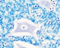
Brain Histology
Overview Cortex (mouse)
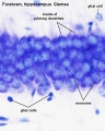
Cortex (mouse)
- Brain histology 03.jpg
- Brain histology 04.jpg


Developing Brain Histology

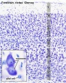
- Human embryo developing cortex, Week 8, Carnegie stage 22.
- Inset (upper right) shows section overview and approximate level of section (red line).
- Thin layer outer called cortical plate will eventually form the adult brain cortex.
- Other underlying layers are part of the development process and will continue to supply cells to the cortex through fetal period, these layers will eventually be almost completely lost.
- Developing cerebrum layer thicknesses are shown in microns.
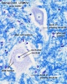
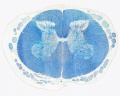
Spinal Cord Histology
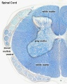
Overview
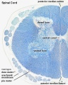
Overview
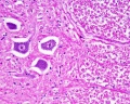
Grey matter
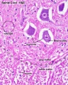
Grey matter (HE)
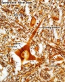
Grey matter (silver)
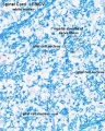
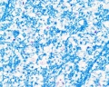
White matter
Overview unlabeled
Grey matter unlabeled 1
Grey matter unlabeled 2
White matter unlabeled 1
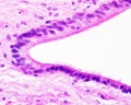
Ependymal cells unlabeled


- Spinal Cord: Overview 1 | Overview 2 | Overview animation | Grey matter | Grey matter | Grey matter | White matter | Overview unlabeled | Grey matter unlabeled 1 | Grey matter unlabeled 2 | White matter unlabeled 1 | Ependymal cells unlabeled
Terms
- artifact - changes and distortions introduced to the normal tissue structure by the histological processing. Common artifacts include: folds (gives the tissue a darker appearance), tears (rips in the tissue can be seen in epithelia), shrinkage when tissues loose mainly liquid through histological processing, and cuts often used in tissue preparation.
Glossary Links
- Glossary: A | B | C | D | E | F | G | H | I | J | K | L | M | N | O | P | Q | R | S | T | U | V | W | X | Y | Z | Numbers | Symbols | Term Link
Cite this page: Hill, M.A. (2024, April 18) Embryology AE Practical - Neural Histology. Retrieved from https://embryology.med.unsw.edu.au/embryology/index.php/AE_Practical_-_Neural_Histology
- © Dr Mark Hill 2024, UNSW Embryology ISBN: 978 0 7334 2609 4 - UNSW CRICOS Provider Code No. 00098G