2012 Group Project 1: Difference between revisions
No edit summary |
No edit summary |
||
| Line 48: | Line 48: | ||
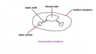
Figure 2 now shows the optic vesicle at slightly later stage in the same simplified cross-section of the embryo, as it migrates from the neural tube to the surface ectoderm. Note the presence of the optic stalk which links the optic vesicle to the neural tube. Later in development, this primitive structure will become the optic nerve, which will link the eye to the brain. | Figure 2 now shows the optic vesicle at slightly later stage in the same simplified cross-section of the embryo, as it migrates from the neural tube to the surface ectoderm. Note the presence of the optic stalk which links the optic vesicle to the neural tube. Later in development, this primitive structure will become the optic nerve, which will link the eye to the brain. | ||
The nerve fibres themselves will initially originate from the retinal ganglion cells in the eye during week 6. After two weeks, these fibers will have grown along the inner wall of the optic stalk and have reached the brain. They grow both in length and width, with the nerve fibres filling the hollow optic stalk to form the solid optic nerve. More than one million nerve fibers will eventually make up the optic nerve, along with glial cells which arise from the inner wall of the optic stalk itself. Myelinisation of the optic nerve begins much later in development at around 7 months, beginning at the optic chiasm and moving towards the eye. The optic chiasm forms just before the nerves reach the brain, and is where half the nerve fibres from each eye will cross over to the opposite side of the brain. | The nerve fibres themselves will initially originate from the retinal ganglion cells in the eye during week 6.<ref>http://www.mdconsult.com/books/page.do?eid=4-u1.0-B978-0-443-06811-9..10017-X--sec8&isbn=978-0-443-06811-9&uniqId=360885019-2#4-u1.0-B978-0-443-06811-9..10017-X--fig17</ref> After two weeks, these fibers will have grown along the inner wall of the optic stalk and have reached the brain. They grow both in length and width, with the nerve fibres filling the hollow optic stalk to form the solid optic nerve. More than one million nerve fibers will eventually make up the optic nerve, along with glial cells which arise from the inner wall of the optic stalk itself. Myelinisation of the optic nerve begins much later in development at around 7 months, beginning at the optic chiasm and moving towards the eye. The optic chiasm forms just before the nerves reach the brain, and is where half the nerve fibres from each eye will cross over to the opposite side of the brain. | ||
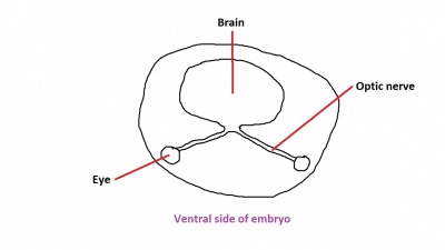
[[File:Formation of the optic nerve and chiasm 1.jpg|400px|thumb|center|Fig. 3: A recognisable brain and eye structure in later development.]] | [[File:Formation of the optic nerve and chiasm 1.jpg|400px|thumb|center|Fig. 3: A recognisable brain and eye structure in later development.]] | ||
Figure 3 demonstrates this. Note the crossing over of the optic nerves just before they enter the brain, at the optic chiasm. This organisation is now much more familiar, with the eyes near the ectoderm and the optic nerve leading through the mesoderm to the brain buried deep in the embryo. | |||
Revision as of 15:56, 10 September 2012
Vision Development
--Mark Hill 12:20, 15 August 2012 (EST) This is a better title.
Introduction
Eyes are an important sensory organ shared across many different species and allow organisms to gather useful visual information from their environment. The visual system uses light from the environment and processes this information in the brain for visual perception. The visual system is complex, and is made up of various structures that work together to form vision. Each of the structures in the eye have specific tasks which contribute to the visual system.
The main anatomical structures of the eye are as follows:
-Cornea
-Sclera
-Iris
-Ciliary body
-Choroid
-Retina
-Anterior chamber
-Posterior chamber
Research history
Development
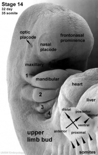
The eye itself is formed from three main components; the optic placode of the head ectoderm, the optic vesicle of the mesoderm (lying under the optic placode) and the cells of the developing brain from the neural tube. The optic placode contributes the lens to the eye, the optic vesicle gives rise to the cornea and sclera, while the neural tube will produce the retina and choroid layers.[1] Cells from the neural tube will also produce the optic nerve, which receives nerve impulses from the retina of the eye. Eyes initially form as laterally paired structures and migrate medially in the human embryo. In other animals such as birds and lizards, the eyes do not migrate and develop laterally on the head. The optic placodes become prominent on the surface of the embryo at approximately Stage 14 of development.
Optic Nerve
The optic nerve consists of nerve fibres that transmit information from the retinal photoreceptor cells to the brain. The optic nerve is formed from the optic stalk. The optic stalk develops as the optic vesicle migrates from its origin in the neural tube to its destination - the surface ectoderm - where it will fuse with the optic placode (also known as the lens placode, which will contribute the lens to the eye).
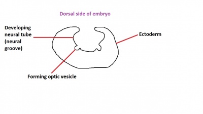
As can be seen in figure 1 above, the optic vesicle forms from the neural tube. However, note that the neural tube has not yet closed, and is still the neural groove at this point.
Figure 2 now shows the optic vesicle at slightly later stage in the same simplified cross-section of the embryo, as it migrates from the neural tube to the surface ectoderm. Note the presence of the optic stalk which links the optic vesicle to the neural tube. Later in development, this primitive structure will become the optic nerve, which will link the eye to the brain.
The nerve fibres themselves will initially originate from the retinal ganglion cells in the eye during week 6.[2] After two weeks, these fibers will have grown along the inner wall of the optic stalk and have reached the brain. They grow both in length and width, with the nerve fibres filling the hollow optic stalk to form the solid optic nerve. More than one million nerve fibers will eventually make up the optic nerve, along with glial cells which arise from the inner wall of the optic stalk itself. Myelinisation of the optic nerve begins much later in development at around 7 months, beginning at the optic chiasm and moving towards the eye. The optic chiasm forms just before the nerves reach the brain, and is where half the nerve fibres from each eye will cross over to the opposite side of the brain.
Figure 3 demonstrates this. Note the crossing over of the optic nerves just before they enter the brain, at the optic chiasm. This organisation is now much more familiar, with the eyes near the ectoderm and the optic nerve leading through the mesoderm to the brain buried deep in the embryo.
Retina
The retinal component of the eye is formed when the optic vesicle fuses with migrating cells from the developing brain.

Ciliary Body
EC
Iris
Lens
The lens has its origin from the optic placode, which develops on the ectodermic surface of the embryo and migrates both medially and inwards into the embryo. The lens allows accommodation of the eye, and adjusts its thickness in order to focus on near or far objects.

Aqueous Chambers
Cornea
Choroid and Sclera
Eyelids
The eyelids are ectodermal in origin and are an extension of the skin which covers and protects the eye.
Lacrimal Glands
Extraocular muscles
Current research
Useful links
Glossary
Image gallery
References
- ↑ http://www.vetmed.vt.edu/education/curriculum/vm8054/eye/EMBYEYE.HTM
- ↑ http://www.mdconsult.com/books/page.do?eid=4-u1.0-B978-0-443-06811-9..10017-X--sec8&isbn=978-0-443-06811-9&uniqId=360885019-2#4-u1.0-B978-0-443-06811-9..10017-X--fig17
- ↑ http://embryology.med.unsw.edu.au/embryology/index.php?title=ANAT2341_Lab_6_-_Early_Embryo
- ↑ http://embryology.med.unsw.edu.au/embryology/index.php?title=ANAT2341_Lab_6_-_Early_Embryo
External Links
External Links Notice - The dynamic nature of the internet may mean that some of these listed links may no longer function. If the link no longer works search the web with the link text or name. Links to any external commercial sites are provided for information purposes only and should never be considered an endorsement. UNSW Embryology is provided as an educational resource with no clinical information or commercial affiliation.
--Mark Hill 12:22, 15 August 2012 (EST) Please leave the content listed below the line at the bottom of your project page.
2012 Projects: Vision | Somatosensory | Taste | Olfaction | Abnormal Vision | Hearing


