2011 Group Project 1
| Note - This page is an undergraduate science embryology student group project 2011. |
Turner Syndrome
Introduction
Turner syndrome (or Ullrich-Turner syndrome) is one of the most common chromosomal disorders, caused by complete or partial X monosomy in some or all cells. Approximately 1 in 2000 female live births, however the morbidity rate of spontaneous abortions is 10% and only about 1% of fetuses survive to term.
During normal fetal development, ovaries contain as many as 7 million oocytes. The oocytes gradually reduced to 400,000 during menarche and during menopause fewer than 10,000 remains. However, in Turner syndrome, the ovaries develop normally during embryogenesis but the absence of the second X chromosome leads to an accelerated loss of oocytes, which is complete by age 2 years. In this case genetically menopause occurs before menarche and the ovaries are reduced to atrophic fibrous strands, devoid of ova and follicles (streak ovaries). What is also affected is the development of somatic (nongonadal) tissues that reside on the missing/abnormal X chromosome. For example short stature is caused by a deletion of the Xp chromosome and the deletion of Xq causes gonadal dysfunction.
The most frequent clinical feature is short stature and gonadal dysgenesis. Other features may include coarctation of the aorta, renal anomalies, neck webbing, and lymphoedema. The affected organ systems and tissues may are effected to a lesser or greater extent amongst that are affected by turner syndrome. A multidisciplinary approach to treatment is important to improve the quality of life of girls with Turner Syndrome.
History
Epidemiology
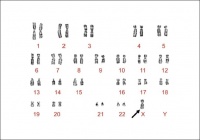
Turner Syndrome affects about 1 in 2000 live-born females. There are three types of karyotypic abnormalities but the most frequently seen is where the entire X chromosome is missing resulting in 45 X karyotype. The remaining third have structural abnormalities of the X chromosomes, and two thirds are mosaics. Whereby, the maternal X is retained in two-thirds of women and the paternal X in the remainder. The phenotype of Turner Syndrome is varies but it involves anomalies of the sex chromosome. It could be caused by the limited amount of genetic material in these abnormal chromosomes. Turner Syndrome can be transmitted from mother to daughter, and thus can it could be described as a heredity linked syndrome.
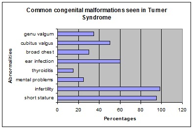
Table 1. Abnormalities associated with Turner Syndrome
Etiology
Clinical manifestations
Diagnostic Procedures
Treatment
The most common abnormality is short stature as bone age is delaged due to the lack of estrogen. This could be treated with the completion of growth hormone therapy.
For young girls use a low dose of natural estrogen to promote and maintain secondary sex characteristics. However recommended to be used when she is approaching puberty in terms of social circumstances as physical sex appearance becomes apparent among her peers.
Current/future research possibilities
Glossary
References
2011 Projects: Turner Syndrome | DiGeorge Syndrome | Klinefelter's Syndrome | Huntington's Disease | Fragile X Syndrome | Tetralogy of Fallot | Angelman Syndrome | Friedreich's Ataxia | Williams-Beuren Syndrome | Duchenne Muscular Dystrolphy | Cleft Palate and Lip

