2010 BGD Practical 6 - Week 3: Difference between revisions
(Created page with '==Introduction== {{Template:2010BGDLab6}} {{Template:2010BGDLab6}} ---- {{Template:BGDFooter2010}}') |
No edit summary |
||
| Line 2: | Line 2: | ||
{{Template:2010BGDLab6}} | {{Template:2010BGDLab6}} | ||
==Introduction== | |||
Key events of human development during the third week (week 3) following fertilization or Clinical week 5 (LMP). Note that during this time the conceptus cells not contributing to the embryo are contributing to placental membranes and the early placenta. | |||
{| class="prettytable" | |||
| <center>'''Day'''</center> | |||
| <center>'''Stage'''</center> | |||
| '''Event''' | |||
|- | |||
| <center>15</center> | |||
| | |||
| | |||
|- | |||
| <center>16</center> | |||
| [[Carnegie stage 7|Stage 7]] | |||
| [[Image:Stage7.jpg|120px|left]] '''Primitive node''' (Hensen's node, primitive knot) The small circular region located at the cranial end of the primitive streak, where gastrulation occurs, and is a controller of this process. The second role is to act as an initial generator of the left-right (L-R) body axis. | |||
|- | |||
| <center>17</center> | |||
| | |||
| | |||
|- | |||
| <center>18</center> | |||
| [[Carnegie stage 8|Stage 8]] | |||
| [[Image:Stage8_human.jpg|120px|left]] [http://embryology.med.unsw.edu.au/Notes/neuron.htm Neural] neurogenesis, neural groove and folds are first seen | |||
|- | |||
| <center>19</center> | |||
| | |||
| [[Image:Stage8_SEM1.jpg|120px|left]] | |||
|- | |||
| <center>20</center> | |||
| [[Carnegie stage 9|Stage 9]] | |||
| [[File:Stage9_bf1c.jpg|200px|link=Carnegie stage 9]] [http://embryology.med.unsw.edu.au/Notes/skmus6.htm Musculoskeletal] somitogenesis, first somites form and continue to be added in sequence caudally | |||
[http://embryology.med.unsw.edu.au/Notes/neuron.htm Neural] the three main divisions of the brain, which are not cerebral vesicles, can be distinguished while the neural groove is still completely open | |||
[http://embryology.med.unsw.edu.au/Notes/ncrest.htm Neural Crest] mesencephalic neural crest is visible [http://www.ncbi.nlm.nih.gov/pubmed/17848161 PMID: 17848161] | |||
|- | |||
| <center>21</center> | |||
| | |||
| [http://embryology.med.unsw.edu.au/Notes/heart.htm Heart] cardiogenesis, week 3 begins as paired heart tubes. | |||
|} | |||
==Week 2 and 3 Movies== | |||
{| border='0px' | |||
|- | |||
| [[File:Week2_001 icon.jpg|90px|link=Development_Animation_-_Implantation]] | |||
| [[File:Mesoderm 001 icon.jpg|90px|link=Development_Animation_-_Mesoderm]] | |||
| [[File:Chorion 001 icon.jpg|90px|link=Development Animation - Chorionic Cavity]] | |||
| [[File:Amnion 001 icon.jpg|90px|link=Development Animation - Amniotic Cavity]] | |||
| [[File:Week3_folding icon.jpg|90px|link=Development Animation - Week 3]] | |||
|- | |||
| [[Development_Animation_-_Implantation|Implantation]] | |||
| [[Development_Animation_-_Mesoderm|Mesoderm]] | |||
| [[Development Animation - Chorionic Cavity|Chorionic Cavity]] | |||
| [[Development Animation - Amniotic Cavity|Amniotic Cavity]] | |||
| [[Development_Animation_-_Week 3|Week 3]] | |||
|- | |||
|} | |||
| Line 9: | Line 76: | ||
{{Template:BGDFooter2010}} | {{Template:BGDFooter2010}} | ||
[[Category:Week 3]] | |||
Revision as of 15:20, 13 May 2010
Introduction
Practical 6: Week 3 | Week 4 | Week 5 | Week 6 | Week 7 | Week 8 | Quiz
Introduction
Key events of human development during the third week (week 3) following fertilization or Clinical week 5 (LMP). Note that during this time the conceptus cells not contributing to the embryo are contributing to placental membranes and the early placenta.
| Event | ||
| Stage 7 |  | |
| Stage 8 |  | |
 | ||
| Stage 9 | 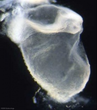 Musculoskeletal somitogenesis, first somites form and continue to be added in sequence caudally Musculoskeletal somitogenesis, first somites form and continue to be added in sequence caudally
Neural the three main divisions of the brain, which are not cerebral vesicles, can be distinguished while the neural groove is still completely open Neural Crest mesencephalic neural crest is visible PMID: 17848161 | |
| Heart cardiogenesis, week 3 begins as paired heart tubes. |
Week 2 and 3 Movies
| Implantation | Mesoderm | Chorionic Cavity | Amniotic Cavity | Week 3 |
Practical 6: Week 3 | Week 4 | Week 5 | Week 6 | Week 7 | Week 8 | Quiz
Glossary Links
- Glossary: A | B | C | D | E | F | G | H | I | J | K | L | M | N | O | P | Q | R | S | T | U | V | W | X | Y | Z | Numbers | Symbols | Term Link
- 2010 BGD: Lecture 1 | Lecture 2 | Practical 3 | Practical 6 | Practical 12
Cite this page: Hill, M.A. (2024, April 25) Embryology 2010 BGD Practical 6 - Week 3. Retrieved from https://embryology.med.unsw.edu.au/embryology/index.php/2010_BGD_Practical_6_-_Week_3
- © Dr Mark Hill 2024, UNSW Embryology ISBN: 978 0 7334 2609 4 - UNSW CRICOS Provider Code No. 00098G