2010 BGD Practical 3 - Notochord
Practical 3: Oogenesis and Ovulation | Gametogenesis | Fertilization | Early Cell Division | Week 1 | Implantation | Week 2 | Extraembryonic Spaces | Gastrulation | Notochord | Week 3 | Quiz
Introduction
The embryonic structure which establishes body axes and patterns surrounding tissues is called the notochord.
The notochord is a midline column of cells running in a rostrocaudal direction (head-tail) within the mesoderm layer. It exists as a transient developmental patterning structure with a role in molecular signaling (patterning) and controlling the direction of embryonic disc folding (mechanical).
The notochordal process begins as a fold of ectoderm extending cranially toward the prechordal plate region. The sequence of differentiation: notochordal process -> notochordal plate -> notochord.
- Elongation of the notochordal process cranially from the primitive pit as a hollow tube (notochordal canal) in the midline of the embryonic disc underlying the ectoderm.
- The notochordal canal may appear to break down on the endodermal side forming a notochordal plate continuous with the endodermal layer.
- Notochordal plate folds to form notochord. The notochord (also called axial mesoderm) is an embryonic structure that regulates differentiation of surrounding structures including the overlying ectoderm (neural plate) and mesoderm (somites).
Practical 3: Oogenesis and Ovulation | Gametogenesis | Fertilization | Early Cell Division | Week 1 | Implantation | Week 2 | Extraembryonic Spaces | Gastrulation | Notochord | Week 3 | Quiz
| The embryonic structure which establishes body axes and patterns surrounding tissues is called the notochord.
The notochord is a midline column of cells running in a rostrocaudal direction (head-tail) within the mesoderm layer. It exists as a transient developmental patterning structure with a role in molecular signaling (patterning) and controlling the direction of embryonic disc folding (mechanical). |
File:Stage7n3.jpgFile:Image 001.gif
|
Disc Folding
Folding: all edges of the embryonic disc will fold ventrally, forming a rostro-caudal "C" shaped tube.
Notochord Development
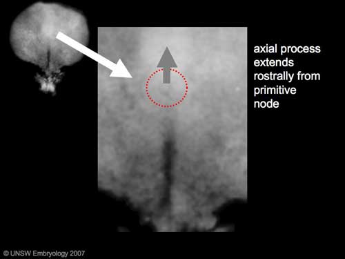
Formation of the axial process which then forms the notochord (also called axial mesoderm). This mesodermal column of cells determines the direction of embryonic disc folding and signals the differentiation of nearby mesoderm (somites) and overlying ectoderm (neural plate then tube).
The notochordal process begins as a fold of ectoderm extending cranially toward the prechordal plate region. The sequence of differentiation: notochordal process -> notochordal plate -> notochord.
(a) Elongation of the notochordal process cranially from the primitive pit as a hollow tube (notochordal canal) in the midline of the embryonic disc underlying the ectoderm.
(b) The notochordal canal may appear to break down on the endodermal side forming a notochordal plate continuous with the endodermal layer.
(c) Notochordal plate folds to form notochord. The notochord (also called axial mesoderm) is an embryonic structure that regulates differentiation of surrounding structures including the overlying ectoderm (neural plate) and mesoderm (somites).
(figures- Moore and Persaud, 1998)
Glossary Links
- Glossary: A | B | C | D | E | F | G | H | I | J | K | L | M | N | O | P | Q | R | S | T | U | V | W | X | Y | Z | Numbers | Symbols | Term Link
- 2010 BGD: Lecture 1 | Lecture 2 | Practical 3 | Practical 6 | Practical 12
Cite this page: Hill, M.A. (2024, April 19) Embryology 2010 BGD Practical 3 - Notochord. Retrieved from https://embryology.med.unsw.edu.au/embryology/index.php/2010_BGD_Practical_3_-_Notochord
- © Dr Mark Hill 2024, UNSW Embryology ISBN: 978 0 7334 2609 4 - UNSW CRICOS Provider Code No. 00098G
Terms
- bilaminar- having 2 layers
- blastocyst- the developmental stage following morula, as this stage matures, the zona pellucia is lost allowing the conceptus to adplant and then implant into the uterine wall.
- inner cell mass- the clump of cells found inside the blastocyst. These cells will go in to form the embryo, these are the "stem cells" (we here about in the media) that are totipotential, they can form any tissue in the embryo. Mature oocyte-the female germ cell released at ovulation from the ovary.
- parental genomes- the male (sperm) and female (oocyte) DNA which contributes to the embryo's cells.
- trilaminar embryonic disc- the 3 layered embryo stage.
- Trophoblasts- (Gr. trophe = nutrition) outer layer of cells on blastocyst that will generate the embryonic part of the placenta.
Glossary Links
- Glossary: A | B | C | D | E | F | G | H | I | J | K | L | M | N | O | P | Q | R | S | T | U | V | W | X | Y | Z | Numbers | Symbols | Term Link
- 2010 BGD: Lecture 1 | Lecture 2 | Practical 3 | Practical 6 | Practical 12
Cite this page: Hill, M.A. (2024, April 19) Embryology 2010 BGD Practical 3 - Notochord. Retrieved from https://embryology.med.unsw.edu.au/embryology/index.php/2010_BGD_Practical_3_-_Notochord
- © Dr Mark Hill 2024, UNSW Embryology ISBN: 978 0 7334 2609 4 - UNSW CRICOS Provider Code No. 00098G

