2010 BGD Practical 3 - Extraembryonic Spaces
Practical 3: Oogenesis and Ovulation | Gametogenesis | Fertilization | Early Cell Division | Week 1 | Implantation | Week 2 | Extraembryonic Spaces | Gastrulation | Notochord | Week 3 | Quiz
Introduction
Practical Audio
 |
BGD Cycle A 2010 Audio - Dr Mark Hill Monday 10th May 2010 3-5pm G2G4. (this class was interrupted by a server crash)
listen Part 5 | download (1.32 Mb MP3 11:33) |

The conceptus is now fully implanted witin the endometrial wall and the uterine epithelium has reformed over the site of implanation.
Note that all future development will now occur within the wall of the uterus, not within the cavity of the uterine body.
As the conceptus continues to implant and grow a series of fluid-filled cavities form both outside and inside the implanting conceptus.
- Outside the Conceptus - maternal blood-filled lacunae
- Inside the Conceptus - 3 separate spaces - amniotic sac, yolk sac, chorionic sac
Conceptus Cavities Week 2 and Week 3
Stage 7 showing the chorion and yolk sac
Stage 7 higher power of embryonic disc and yolk sac
Stage 9


Outside
Continued expansion of syncitiotrophoblasts within the endometrial wall opens both uterine glands and uterine blood vessels. These spaces outside the conceptus fill with uterine gland secretions and maternal blood forming maternal blood-filled lacunae. These lacunae provide the initial nutrition to the growing conceptus, which will later be provided through the placenta.
Inside
A cavity forms now between the inner cell mass and the trophoblast (cytotrophoblasts) wall. This cavity is the amniotic cavity.
A new cell layer forms from cells proliferating and lining the inside of the blastoceol cytotrophoblastic layer (extraembryoic mesoderm). This extraembryoic mesoderm layer continues to proliferate and then vacuolates, splitting into two separate cavities.
Overview of blastocyst implantation in uterine wall during the second week of development. (Image: Moore & Persaud, 1998)
Beginning of Carnegie Stage 6, blastocyst fully implanted in endometrial wall.
Continued expansion of the cavities in the extraembryonic mesoderm, allow the primary yolk sac to collapse. The collapsed space later becomes lined with endodermal cells and forms the yolk sac.
The space formed outside is lined with extraembryonic mesoderm and forms the third cavity the chorionic cavity.
A section of extraembryonic mesoderm links the now bilaminar embryonic disc to surrounding shell and is called the connecting stalk.
Human Conceptus Implantation 2nd and 3rd Week of Development
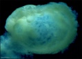
| Identify chorionic sac, yolk sac and amniotic sac, all containing different fluids.
Identify secondary chorionic villi, maternal blood "spaces" (empty) and uterine tissue (maternal decidua). The embryo lies between the yolk sac fluid and the amniotic sac fluid. Note that the ends ("bases") of some long secondary chorionic villi are attached to the maternal (decidual) tissue by dense clusters of trophoblast cells (the cytotrophoblastic column) - these are anchoring secondary villi. Other shorter, secondary villi are "free-floating" in the maternal intervillous spaces, from which the blood has drained into the large endometrial veins. (Image: Nishimura etal., 1977) |
 Carnegie Stage 6-7, human conceptus approximately 16-18 days. (about the time of the first missed menstrual period). Carnegie Stage 6-7, human conceptus approximately 16-18 days. (about the time of the first missed menstrual period).
|
--Mark Hill 15:34, 16 June 2010 (UTC) These 2 missing images Carnegie Stage 6-7 from the original practical class page have been added later and are not examinable.
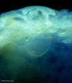
Human Conceptus (high power view)
| Identify the three fluid-filled sacs, the embryo, and the body (connecting) stalk indicating the caudal end of the embryo. Note the thin amnion and the thicker wall of the yolk sac and conversely, the thick ectoderm and thin endoderm. Vascular channels in connecting stalk. The broad elevation in the ectoderm is the expansion dome (primitive single brain bulge) and the cluster of cells at the caudal end of the embryonic disc is the primitive node region. Note the precipitated protein in the extra-embryonic coelom and in the yolk sac, and the start of the formation of the intra-embryonic coelom between ectoderm and entoderm, at the cranial (rostral) end of the expansion dome. | 
|
--Mark Hill 15:34, 16 June 2010 (UTC) These 2 missing Carnegie Stage 6-7 images from the original practical class page have been added later and are not examinable.
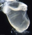
Human Conceptus (high power view)

|
Approximate cross-section of an 18 day human conceptus.
(Image: Nishimura etal., 1977)
|
Practical 3: Oogenesis and Ovulation | Gametogenesis | Fertilization | Early Cell Division | Week 1 | Implantation | Week 2 | Extraembryonic Spaces | Gastrulation | Notochord | Week 3 | Quiz
Terms
- bilaminar- having 2 layers
- blastocyst- the developmental stage following morula, as this stage matures, the zona pellucia is lost allowing the conceptus to adplant and then implant into the uterine wall.
- inner cell mass- the clump of cells found inside the blastocyst. These cells will go in to form the embryo, these are the "stem cells" (we here about in the media) that are totipotential, they can form any tissue in the embryo. Mature oocyte-the female germ cell released at ovulation from the ovary.
- parental genomes- the male (sperm) and female (oocyte) DNA which contributes to the embryo's cells.
- trilaminar embryonic disc- the 3 layered embryo stage.
- trophoblasts- (Gr. trophe = nutrition) outer layer of cells on blastocyst that will generate the embryonic part of the placenta.
Glossary Links
- Glossary: A | B | C | D | E | F | G | H | I | J | K | L | M | N | O | P | Q | R | S | T | U | V | W | X | Y | Z | Numbers | Symbols | Term Link
- 2010 BGD: Lecture 1 | Lecture 2 | Practical 3 | Practical 6 | Practical 12
Cite this page: Hill, M.A. (2024, April 19) Embryology 2010 BGD Practical 3 - Extraembryonic Spaces. Retrieved from https://embryology.med.unsw.edu.au/embryology/index.php/2010_BGD_Practical_3_-_Extraembryonic_Spaces
- © Dr Mark Hill 2024, UNSW Embryology ISBN: 978 0 7334 2609 4 - UNSW CRICOS Provider Code No. 00098G