2009 Lecture 16
Genital Development
Introduction
This section of notes covers genital development. Differences in development are dependent on a protein product of the Y chromosome SRY gene. Mesonephric duct (Wolffian Duct) and paramesonephric (Mullerian Duct) contribute the majority of male and female internal genital tract respectively.
Objectives
- Understand the role of the Y chromosome in sex determination.
- Understand the differences in male/female duct develpoment (mesonephric/paramesonephric).
- Compare the development of the cloaca in the male and female.
- Understand the developmental abnormalities in male and female development.
Textbooks
- Human Embryology (2nd ed.) Larson Ch10 p261-306
- The Developing Human: Clinically Oriented Embryology (6th ed.) Moore and Persaud Chapter 13 p303-346
Three Stages
The mesonephric/paramesonephric duct changes are one of the first male/female differences that occur in development, while external genitaila remain indeterminate in appearance for quite a while.
- Differentiation of gonad (Sex determination)
- Differentiation of internal genital organs
- Differentiation of external genital organs
The 2nd and 3rd stages dependent on endocrine gonad. Reproductive development has a long maturation timecourse, begining in the embryo and finishing in puberty. (More? Puberty)
Development Overview
Sex Determination
- Humans (week 5-6)
- Germ cells migrate into gonadal ridge
- Gonads (male/female) identical at this stage, indifferent
Gonad development
- dependent on sex chromosome
- Y testes
- No Y ovary
SRY
- SRY protein (Testes determining factor, TDF) binds DNA
- Transcription factor, Bends DNA 70-80 degrees
Internal Genital Organs
- All embryos form paired
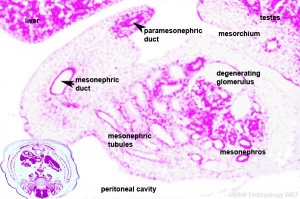
- Mesonephric duct, see kidney development
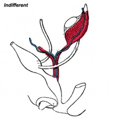
- Paramesonephric duct, Humans 7th week Invagination of coelomic epithelium Cord grows and terminates on urogenital sinus
- Male Gonad (testes) secretes Mullerian duct inhibitory factor (MDIF) which causes regression of paramesonephric duct
- Male Gonad (testes) secretes Testosterone which retains mesonephric duct
External Genital Organs
- All embryos initially same (indifferent)
- Testosterone differentiates male
Human Timeline
- 24 days - intermediate mesoderm, pronephros primordium
- 28 days - mesonephros and mesonephric duct
- 35 days - uteric bud, metanephros, urogenital ridge
- 42 days - cloacal divison, gonadal primordium (indifferent)
- 49 days - paramesonephric duct, gonadal differentiation
- 56 days - paramesonephric duct fusion (female)
- 100 days - primary follicles (ovary)
Kidney
- kidneys and genital ridge develop from intermediate mesoderm, which lies between the lateral plate mesoderm and the somites.
- kidney develops in multiple stages, which occur in a rostrocaudal sequence.
- earliest structure to form is the pronephros, in week 4, featuring a pronephric duct with associated nephrogenic mesenchyme.
- pronephros degenerates early on, leaving only the duct system running down to the cloaca – this becomes known as the mesonephric duct, in the embryo.
- next stage is the formation of the mesonephros, also in week 4. Its differentiation is induced by the pronephros.
- mesonephros is also a transient structure. It provides a template for the adult metanephros, beginning on day 35-37.
Sex Determination
Male (XY)
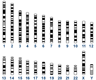
- Y chromosome - 200+ genes, 50 million base pairs
- Sry was discovered (1990) by studying a human XY female, resulting from a deletion in the Y chromosome that did not allow testis development. Subsequent mapping of this deletion allowed isolation and characterization of the SRY gene.
- SRY encodes: encodes a 204 amino acid protein (Mr 23884 Da) that is a zinc-finger transcription factor. Transcription factors bind to specific sites of DNA and regulates the transcription (expression) of other genes, we still do not know all the genes SRY regulates.
- SRY is expressed: when testes begin to form, in gonadal tissue and does not require the presence of germ cells.
- induces cells to differentiate into Sertoli cells
Sertoli cells
- produce signals that promote development of male characteristics
- suppress development of female characteristics
- induce primordial germ cells to commit to sperm development
Female (XX)
- 1400+ genes, 150 million base pairs
- In contrast to the Y chromosome, the X chromosome contains about 5% of the haploid genome and encodes house-keeping and specialized functions. The genetic content of the X chromosome has been strongly conserved between species.
- Genes such as Wnt-4 and DAX-1 necessary for initiation of female pathway ovary development
- female not considered a default process
- An early discovery (1961) was that in order to have correct levels of X chromosome gene/protein expression (gene dosage), females must "inactivate" a single copy of the X chromosome in each and every cell. The initiator of the X inactivation process was discovered (1991) to be regulated by a region on the inactivating X chromosome encoding an X inactive specific transcript (XIST), that acts as RNA and does not encode a protein.
- X inactivation occurs randomly throughout the embryo, generating a mosaic of maternal and paternally derived X chromosome activity in all tissues and organs.
Primordial Germ Cells
- thought to be the first population of cells to migrate through the primitive streak in early gastrulation.
- This population of cells then lie at the hindgut yolk sac junctional region and later migrate into the germinal ridge in early embryonic development.
- It is not the primordial germ cells which respond to SRY presence or absence, but the supporting cells within the developing gonad.
Gametogenesis
- forming PGCs and getting them into genital ridge as gonad forms
- formation of germ plasm and determination of PGCs
- migration of PGCs into developing gonads
- process of meiosis and modifications of meiosis for forming sperm and eggs
- differentiation of sperm and egg
- hormonal control of gamete maturation and ovulation
Male Gonadal Development
This looped animation shows the development of the male gonad showing medullary sex cords.
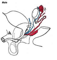
The paramesonephric duct (red, left) degenerates under the influence of Mullerian duct inhibitory factor (MDIF) secreted by sertoli cells (differentiated by SRY expression).
The mesonephric duct (purple) differentiates under the influence of Testosterone secreted by Leydig cells. Within the testes these mesonephric tubules grow towards the medullary sex cords and will form the rete teste. The mesonephric duct extending out of the gonad forms the ductus deferens.
The medullary sex cords (orange) form testis cords that later differentiate into solid seminiferous tubules which become hollow and actively produce spermatazoa during puberty.
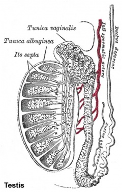
The tunica albuginea (white) covers the testis and bands extend inward to form connective tissue septa.
Female Gonadal Development
This looped animation shows the development of the female gonad showing cortical sex cords.
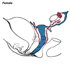
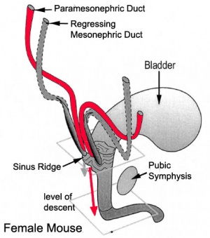
The mesonephric duct (purple) degenerates, small remnants may remain as epoophoron and paroophoron (in the mesentry of the ovary) and Gartner's cycts (near vagina).
The paramesonephric duct (red, left) grows forming the oviduct (uterine horn) and the end opens into the peritoneal cavity and terminates in fimbria (finger-like extensions). Away from the ovary, the two paramesonephric ducts fuse in the midline to form the uterus.
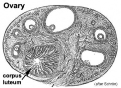
The cortical sex cords (orange) form after the primary sex cords degenerate and mesothelium forms secondary cords. The surrounding connective tissue (pink) differentiates to form follicle cells.
Fetal
- late embryonic male genital development and now in fetal development we will firstly observe early fetal female development.
References
- Before We Are Born (5th ed.) Moore and Persaud Chapter 14 p289-326
- Essentials of Human Embryology, Larson Ch10 p173-205
- Human Embryology, Fitzgerald and Fitzgerald Ch21-22 p134-152
- Developmental Biology (6th ed.) Gilbert Ch14 Intermediate Mesoderm
Online Links
- UNSW Embryology Abnormalities | Y chromosome | X chromosome | Ovary | Stage 13/14 Embryo | Stage 22 Embryo | Stage 22 Highpower
- UNSW Embryology Movies: Urogenital Movies
- Embryo Images Unit: Embryo Images Online | Urongenital Development | Internal Genitalia | Definitive Kidney | External Genitalia
- Histology: Male Reproductive System | Female Reproductive System
Glossary Links
- Glossary: A | B | C | D | E | F | G | H | I | J | K | L | M | N | O | P | Q | R | S | T | U | V | W | X | Y | Z | Numbers | Symbols | Term Link
Course Content 2009
Embryology Introduction | Cell Division/Fertilization | Cell Division/Fertilization | Week 1&2 Development | Week 3 Development | Lab 2 | Mesoderm Development | Ectoderm, Early Neural, Neural Crest | Lab 3 | Early Vascular Development | Placenta | Lab 4 | Endoderm, Early Gastrointestinal | Respiratory Development | Lab 5 | Head Development | Neural Crest Development | Lab 6 | Musculoskeletal Development | Limb Development | Lab 7 | Kidney | Genital | Lab 8 | Sensory - Ear | Integumentary | Lab 9 | Sensory - Eye | Endocrine | Lab 10 | Late Vascular Development | Fetal | Lab 11 | Birth, Postnatal | Revision | Lab 12 | Lecture Audio | Course Timetable
Cite this page: Hill, M.A. (2024, April 18) Embryology 2009 Lecture 16. Retrieved from https://embryology.med.unsw.edu.au/embryology/index.php/2009_Lecture_16
- © Dr Mark Hill 2024, UNSW Embryology ISBN: 978 0 7334 2609 4 - UNSW CRICOS Provider Code No. 00098G
