1987 Developmental Stages In Human Embryos - Stage 21
| Embryology - 25 Apr 2024 |
|---|
| Google Translate - select your language from the list shown below (this will open a new external page) |
|
العربية | català | 中文 | 中國傳統的 | français | Deutsche | עִברִית | हिंदी | bahasa Indonesia | italiano | 日本語 | 한국어 | မြန်မာ | Pilipino | Polskie | português | ਪੰਜਾਬੀ ਦੇ | Română | русский | Español | Swahili | Svensk | ไทย | Türkçe | اردو | ייִדיש | Tiếng Việt These external translations are automated and may not be accurate. (More? About Translations) |
O'Rahilly R. and Müller F. Developmental Stages in Human Embryos. Contrib. Embryol., Carnegie Inst. Wash. 637 (1987).
| Online Editor Note |
|---|
| O'Rahilly R. and Müller F. Developmental Stages in Human Embryos. Contrib. Embryol., Carnegie Inst. Wash. 637 (1987).
The original 1987 publication text, figures and tables have been altered in formatting, addition of internal online links, and links to PubMed. Original Document - Copyright © 1987 Carnegie Institution of Washington.
|
- 1987 Stages: Introduction | 1 | 2 | 3 | 4 | 5 | 6 | 7 | 8 | 9 | 10 | 11 | 12 | 13 | 14 | 15 | 16 | 17 | 18 | 19 | 20 | 21 | 22 | 23 | References | Appendix 1 | Appendix 2 | Historic Papers | Embryonic Development
| Historic Disclaimer - information about historic embryology pages |
|---|
| Pages where the terms "Historic" (textbooks, papers, people, recommendations) appear on this site, and sections within pages where this disclaimer appears, indicate that the content and scientific understanding are specific to the time of publication. This means that while some scientific descriptions are still accurate, the terminology and interpretation of the developmental mechanisms reflect the understanding at the time of original publication and those of the preceding periods, these terms, interpretations and recommendations may not reflect our current scientific understanding. (More? Embryology History | Historic Embryology Papers) |
Stage 21
| Carnegie Collection - Stage 21 | |||||||||||
|---|---|---|---|---|---|---|---|---|---|---|---|
| Serial No. | Size (mm) | Grade | Fixative | Embedding Medium | Plane | Thinness (µm) | Stain | Point Score | Sex | Year | Notes |
| 22 | E, 20 Ch, 35x30x30 | Good | Alc. | P | Transverse | 50 | Al. coch. | 34.5 | Female | 1895 | |
| 57 | E, 23 Ch., ca. 30 | Poor | Alc. | P | Sagittal | 50 | Al. coch. | 36 | Male | 1896 | |
| 128 | E, 20 Ch., 50x43 | Good | Formalin | P | Coronal | 50 | Al. coch. | 33 | Female | 1898 | |
| 229 | E, 19 | Poor | Alc. | P | Sagittal | 50 | Al. coch. | 33 | Female | 1903 | |
| 349 | E, 24 | Good | Zenker | C | Coronal | 250 | Unstained | 36 | ? | 1905 | Double vascular injection |
| 455 | E, 24 Ch., 42x34x20 | Good | Alc. | P | Transverse | 30 | (Stain - Haematoxylin Eosin) | 36.5 | Male | 1910 | |
| 632 | E, 24 Ch., 60x50x30 | Good | Bichlor. acetic | P | Sagittal | 40, 100, 250 | Al. coch. | 33 | Female | 1913 | Injected |
| 903C | E, 23.5 | Good | Formalin | P | Transverse | 40 | Al. coch. | 38.5 | Female | 1914 | |
| 1008 | E, 26,4 | Good | Formalin | P | Sagittal | 40 | Al. coch. | 39 | ?? | 1914 | |
| 1358F | E, 23 | Good | Formalin | P | Sagittal | 40 | Al. coch. | 37.5 | Female | 1916 | |
| 2937 | E,, 24.2 | Good | Bouin | P | Transverse | 50 | (Stain - Haematoxylin Eosin) aur., or. G. | 39 | Female | 1920 | |
| 3167 | E., 24.5 Ch., 60x50x40 | Poor | Bichlor, acetic, formol | P | Transverse | 20 | Al. coch. | 32 | Male | 1920 | |
| 4090 | E, 22.2 Ch.. 66x46x30 | Good | Formalin | P | Transverse | 40 | Al. coch. | 30 | Female | 1922 | |
| 4160 | E,25 | Poor | Formalin | P | Sagittal | 25 | (Stain - Haematoxylin Eosin) | 39 | Male | 1923 | Tubal |
| 4960 | E.22 Ch,, 47x42x28 | Good | Formalin | P | Transverse | 15 | Al. coch., Mallory | 31.5 | Female | 1925 | |
| 5??6 | E. 215 | Good | Formalin | P | Sagittal | 20 | (Stain - Haematoxylin Eosin) | 34 | Female | 1927 | |
| 6531 | E,22 | Poor | Glacial acetic, | C-P | Transverse | 10 | (Stain - Haematoxylin Eosin) | 31.5 | Female | 1931 | Leitz Collection |
| 7254 | E,225 | Exc | Bouin | C-P | Transverse | 20 | (Stain - Haematoxylin Eosin) | 33.5 | Male | 1936 | |
| 7592 | E,22-> | Exc. | Bouin | C-P | Transverse | 20 | (Stain - Haematoxylin Eosin) | 36 | Female | 1937 | |
| 7864 | E., 24 | Exc, | Formalin | C-P | Frontal | 20 | (Stain - Haematoxylin Eosin) | 32.5 | Male | 1941 | |
| 8553 | E., 22 | Exc | Bouin | C-P | Transverse | 12 | (Stain - Haematoxylin Eosin) | 38 | Female | 1947 | |
| 9614 | E,,22 5 | Exc | Bouin | P | Coronal | 10 &15 | Azan | ? | ? | 1958 | Rubella. Hysterectomy |
Abbreviations
| |||||||||||
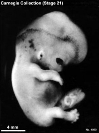
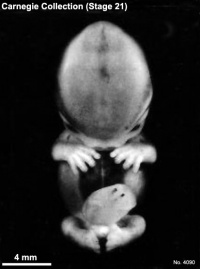
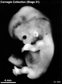
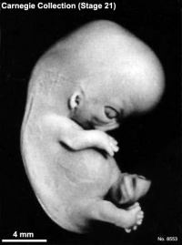
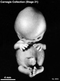
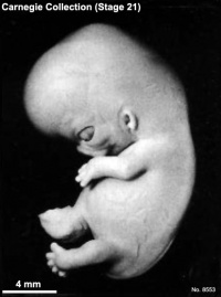
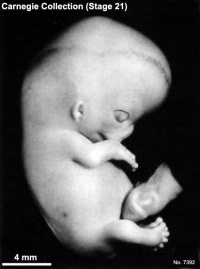
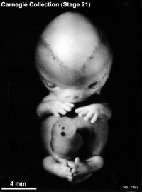
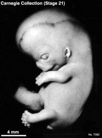
Fig. 21-1. Photographs of three embryos belonging to stage 21. The superficial scalp vascular plexus of the head is plainly visible in several of these views, such as A. It has spread to a level more than halfway between an eye-ear line and the vertex of the head. The fingers are longer and show an early phase in the development of touch pads. These Tastballen are shown at higher magnification by Cummins (1929) at stage 20 (his fig. 7) and stage 22 (his fig. 8.) The hands are flexed at the wrists and are approaching each other over the cardiac region. The lower limbs are curving toward the median plane, and toes of the two feet make contact with each other in some specimens. Top row, No. 4090. Middle row, No. 8553. Bottom row, No 7392. All views are at the same magnification.
Size And Age
Most embryos of this stage measure 22–24 mm.
The age is believed to be approximately 52 postovulatory days.
External Form
The superficial vascular plexus of the head has spread upward to form a line at somewhat more than half the distance from eye-ear level to the vertex.
The fingers are longer and extend further beyond the ventral body wall than they did in the previous stage. The distal phalangeal portions appear slightly swollen and show the beginning of tactile pads. The hands are slightly flexed at the wrists and nearly come together over the cardiac eminence. The feet are also approaching each other, and the toes of the two sides sometimes touch.
Features For Point Scores
- Cornea. Cells are beginning to invade the postepithelial layer, converting it into the substantia propria (Streeter, 1951, fig. 16).
- Optic nerve. Remnants of ependyma are present and may extend along practically the whole length of the optic stalk. A hyaloid groove is visible at the bulbar end. A few nerve fibers are arriving at the brain.
- Cochlear duct. The tip of the duct now points definitely "downward" (fig. 19-6).
- Adenohypophysis. The thread-like stalk is beginning to be absorbed (fig. 19-7).
- Vomeronasal organ. The opening of the sac is reduced in size, a short, narrow neck is present, and the end of the sac is expanded (fig. 19-9).
- Submandibular gland. The duct has begun to form knob-like branches (fig. 19-10).
- Metanephros. Spoon-shaped glomerular capsules are developing, but no large glomeruli are present yet (fig. 19-11).
- Humerus. Cartilaginous phases 1-4 are present (Streeter, 1949, figs. 3, 19, and 20).
Additional Features
- Heart. Some photomicrographs were reproduced by Cooper and O'Rahilly (1971, figs. 15-17).
- Testis. The testis shows a flattened surface epithelium, an underlying tunica albuginea, and branching and anastomosing cords: "the forerunners of the seminiferous tubules" (Wilson, 1926a).
- Brain. A general view of the organ was given by O'Rahilly and Gardner (1971, fig. 1). The olivary nucleus is present in the rhombencephalon. Three-quarters of the surface of the diencephalon is covered by the cerebral hemispheres. The optic tract reaches approximately the site of the lateral geniculate body. The insula can now be recognized as a faint concavity at the surface of the hemisphere.
References
| Online Editor Note |
|---|
| O'Rahilly R. and Müller F. Developmental Stages in Human Embryos. Contrib. Embryol., Carnegie Inst. Wash. 637 (1987).
The original 1987 publication text, figures and tables have been altered in formatting, addition of internal online links, and links to PubMed. Original Document - Copyright © 1987 Carnegie Institution of Washington.
|
See also Müller F & O'Rahilly R. (1990). The human brain at stages 21-23, with particular reference to the cerebral cortical plate and to the development of the cerebellum. Anat. Embryol. , 182, 375-400. PMID: 2252222
Search Pubmed
- 1987 Stages: Introduction | 1 | 2 | 3 | 4 | 5 | 6 | 7 | 8 | 9 | 10 | 11 | 12 | 13 | 14 | 15 | 16 | 17 | 18 | 19 | 20 | 21 | 22 | 23 | References | Appendix 1 | Appendix 2 | Historic Papers | Embryonic Development
| Historic Disclaimer - information about historic embryology pages |
|---|
| Pages where the terms "Historic" (textbooks, papers, people, recommendations) appear on this site, and sections within pages where this disclaimer appears, indicate that the content and scientific understanding are specific to the time of publication. This means that while some scientific descriptions are still accurate, the terminology and interpretation of the developmental mechanisms reflect the understanding at the time of original publication and those of the preceding periods, these terms, interpretations and recommendations may not reflect our current scientific understanding. (More? Embryology History | Historic Embryology Papers) |
Cite this page: Hill, M.A. (2024, April 25) Embryology 1987 Developmental Stages In Human Embryos - Stage 21. Retrieved from https://embryology.med.unsw.edu.au/embryology/index.php/1987_Developmental_Stages_In_Human_Embryos_-_Stage_21
- © Dr Mark Hill 2024, UNSW Embryology ISBN: 978 0 7334 2609 4 - UNSW CRICOS Provider Code No. 00098G

