User:Z5030311
-- Welcome to the 2014 Embryology Course!
- Links: Timetable | How to work online | One page Wiki Reference Card | Moodle
- Each week the individual assessment questions will be displayed in the practical class pages and also added here.
- Copy the assessment items to your own page and provide your answer.
- Note - Some guest assessments may require completion of a worksheet that will be handed in in class with your student name and ID.
| Individual Lab Assessment |
|---|
|
| Lab 12 - Stem Cell Presentation Assessment | More Info | |
|---|---|---|
| Group | Comment | Mark (10) |
| 1/8 |
|
7 |
| 2 |
|
7.5 |
| 3 |
|
7.5 |
| 4 |
|
8.5 |
| 5 |
|
8.5 |
| 6 |
|
8.5 |
| 7 |
|
7.5 |
Z5030311 (talk) 12:45, 6 August 2014 (EST)
Lab Attendance
Lab 1:--Z5030311 (talk) 12:53, 6 August 2014 (EST)
Lab 2:--Z5030311 (talk) 11:21, 13 August 2014 (EST)
Lab 3:--Z5030311 (talk) 11:07, 20 August 2014 (EST)
Lab 4:--Z5030311 (talk) 11:09, 27 August 2014 (EST)
Lab 5:--Z5030311 (talk) 11:12, 3 September 2014 (EST)
Lab 6:--Z5030311 (talk) 11:11, 10 September 2014 (EST)
Lab 7:--Z5030311 (talk) 11:14, 17 September 2014 (EST)
Lab 8:--Z5030311 (talk) 11:07, 24 September 2014 (EST)
Lab 9:--Z5030311 (talk) 11:24, 8 October 2014 (EST)
Lab 10:--Z5030311 (talk) 11:24, 15 October 2014 (EST)
Lab 11:--Z5030311 (talk) 11:17, 22 October 2014 (EST)
Lab 12:--Z5030311 (talk) 11:10, 29 October 2014 (EST)
http://www.ncbi.nlm.nih.gov/pubmed PubMed
Lab 1 Assessment
http://www.ncbi.nlm.nih.gov/pubmed/25036713 <pubmed>25036713</pubmed>
Kisspeptin-54 is essential for human fertility as it is involved in the surge of luteinizing hormone and the maturation of oocytes. Studies have shown that a mutation inactivating the kisspeptin signal leads to infertility in women as there is no surge in the level of luteinizing hormone and so oocytes are not matured and released. In this study 53 women were injected with Kisspeptin-54 following superovulation; it was hoped that the Kisspeptin-54 would cause a surge in LH resulting in oocyte maturation. After 36 hours the oocytes were retrieved transvaginally, their maturation state was assessed and they were fertilized by intracytoplasmic sperm. Embryos were then formed from the fertilized oocyte. It was discovered that an injection of Kisspeptin-54 can increase the mean number of mature eggs produced by each patient and that it can induce oocyte maturation in patients with subfertility who are undergoing in vitro fertilization. In 92% of the patients who were given the Kisspeptin injection the oocyte was fertilized and the subsequent embryo was successfully implanted in the patient’s uterus.
http://www.ncbi.nlm.nih.gov/pubmed/24751928 <pubmed>24751928</pubmed>
One of the stages of IVF is superovulation, this is where multiple oocytes are stimulated to mature by injecting hormones into the patient. This study is proposing to adapt the levels of hormones used in superovulation for each patient so that the optimum number and size of oocytes is achieved. A mathematical model was constructed which predicted the dose of the hormones that would result in the optimum number and size of oocytes. The model was applied to real patients and the resulting oocytes were analyzed to see if the optimum oocytes were produced. The results showed that there were more oocytes and better sized oocytes when the levels of hormones were altered for each patient in comparison to the normal method where the hormone level is the same for each patient. This will improve the success of superovulation cycles and reduce the cost of excess medication.
--Mark Hill These references are related to fertilisation and you have summarised well. (5/5)
Lab 2 Assessment
--Mark Hill The figure legend and reference should have also appeared here on your page. (4/5)
Lab 3 Assessment
Structures that arise from the Ureteric bud
<pubmed>25088264</pubmed> <pubmed>25087982</pubmed>
Structures that arise from the Metanephric mesoderm
<pubmed>18835385</pubmed> <pubmed>19726549</pubmed>
--Mark Hill These reference are relevant. You could have also included a single sentence on why/how you selected these references, including the sub-sections was useful. (5/5)
Lab 4 Assessment
1. <pubmed>24144029</pubmed>
An experimental study of preventing and treating acute radioactive enteritis with human umbilical cordmesenchymal stem cells
Human umbilical cord-derived stem mesenchymal cells were investigated on rats to see if they are able to cure radiation sickness in Humans. The rats used in this experiment had acute radioactive enteritis, which is where there is inflammation of the small intestine. The human stem cells used in the experiment were cultured in vitro and the rat models with the actue radioactive enteritis were established. The stems cells were then injected into the rats and the changes to the Visual and histopathological of the rats were observed.
It was found that rats that were treated with the human umbilical cord-derived stem mesenchymal cells had better survival rates compared to the control group. Histopathologically it was found that the treatment group also had more regenerative cells, stronger proliferation activity and there intestinal mucosa had a better structure.
2. The three developmental vascular "shunts" present in the embryo are Ductus arteriosus, Ductus venosus and Foramen ovale; all three close postnatally.
Ductus arteriosus is a blood vessel which connects the pulmonary artery and the proximal descending aorta; it allows blood to bypass the lungs.
Ductus venosus allows blood from the placenta to bypass the liver by shunting blood from the left umbilical vein to the inferior vena cava.
Foramen ovale is located in the heart and it allows blood to flow from the right atrium to the left atrium; this allows blood to bypass the lungs
--[[User:Z8600021|Mark Hill] Shunts are correct. (4/5)
Lab Assessment 5
Aganglionic colon (Hirschprung's disease)
Hirschprung’s disease is an absence of ganglia in the distal colon causing abnormal function of the gut. The disease is due to an abnormality during the development of the gastrointestinal tract; those individuals with the disease often do not pass meconium in the 24 hours that follow their delivery, patients will also show signs of dysmotility. The disease is diagnosed by histopathological examinations of colon biopsies.
Hirschprung’s disease is caused by the lack of complete migration by neural crest cells during embryonic development, these neural crest cells are precursors of enteric ganglion cells. The lack of enteric ganglion cells in portions of the distal colon result in sections of the colon unable to relax causing obstructions in the colon. As well as intestinal obstructions severe constipation can also occur due to the constant contraction of the gut.
Hirschhprung’s disease can affect different sized portions of the colon, in around 80% of patients only a small portion of the colon is affected and these cases can easily be cured with surgery. However in other patients much larger portions of the colon are affected which makes the cure for the disease harder and can result in death.
<pubmed>24168728</pubmed> <pubmed>25092084</pubmed>
--[[User:Z8600021|Mark Hill] Good summary, you should though spend more time looking for better articles to use. (4/5)
Lab Assessment 7
<pubmed>24227653</pubmed> In this study the effect of the Insm1 gene on the differentiation of endocrine cells in the pituitary gland is investigated. The Insm1 gene encodes a zinc finger transcription factor which is found to be expressed in a variety of endocrine cell types. Mice with an Insm1 mutation were used in this study and it was found that if mice had a mutation in the Insm1 gene then they had an absence or reduced level of a variety of hormones including thyroid-stimulating hormone and growth hormone. The hormones with reduced levels are hormones that are characteristic of the different pituitary cell types. Therefore this study has shown that the Insm1 gene is involved in the differentiation of pituitary cells and that it is vital in embryonic development.
Neural crest-derived mesenchymal cells give rise to the dental papilla as they condense and then invade into the base of the tooth bud. Under the influence of the enamel epithelium the mesenchymal cells also give rise to odontoblasts; the odontoblasts secrete predentin which then calcifies to form dentin.
Another embryonic layer that gives rise to tissues in developing teeth is the ectoderm cells that are present in the oral epithelium, these cells differentiate to become ameloblasts which are cells on the surface of the developing tooth that produce enamel.
In teeth there is a specialised connective tissue that holds the tooth into the bony socket, this structure is called the periodontal ligament.
2. Initial thin ectodermal layer called the dental lamina which overlies the upper and lower jaw bones and this proliferates into two horse-shoe shaped structures which will become the future dental arcades. Enamel organs develop in the dental lamina in the form of rounded swellings and each swelling is the future site of a single tooth. The enamel organ partially encloses the adjacent mesodermal structure known as the dental papilla. Unenclosed mesoderm of the dental papilla surrounds urrounds this and forms a follicular sac. Tooth germ: Enamel organ, dental papilla and follicular sac. Respectively they form the enamel cap of the tooth crown, the dentine and pulp chamber of the tooth and the periodontal membrane.
--[[User:Z8600021|Mark Hill] Excellent (5/5)
Lab Assessment 8
There are several embryonic stages in testis development; these include the differentiation of the gonads into gender specific organs and the differentiation of the internal organs. The differentiation of the external organs and the development of secondary sexual characteristics occur during the foetal and postnatal periods.
In week 6 the genital ridge develops from intermediate mesoderm, this is also known as the undifferentiated gonad; in order for the gonad to differentiate into a specific sex organ signals from particular genes on the Y chromosome are released. The SRY gene, discovered in 1990, is a protein coding gene on the Y chromosome which is essential for the differentiation of the gonads into testes. SRY codes for a 204 amino acid protein; this protein initiates the differentiation of the gonad by binding to specific regions of DNA and expressing certain genes.
SRY causes the primordial of the supporting cells to develop into Sertoli cells, these cells control the further differentiation of the gonad by instructing the germ cells and other cells to go down the male route of development. The Sertoli cells surround the germ cells, form the testis cord and cause Leydig cells to differentiate; this occurs at around day 42. Sertoli cells also express anti-Mullerian hormone (AMH) which prevents female reproductive organs to form, the expression of AMH causes the paramesonephric (Mullerian) duct to degenerate. The Leydig cells produce testosterone and this controls mesonephric (Wolffian) duct differentiation; the rete testis form from the mesonephric tubules that grow towards the testis cords and ductus deferens form from the mesonephric duct extending out of the gonads. The testis cord and the germ cells differentiate into seminiferous tubules. At 49 days there is a clear difference between male and female gonads, after this point the internal organs continue to develop and external organs begin to form.
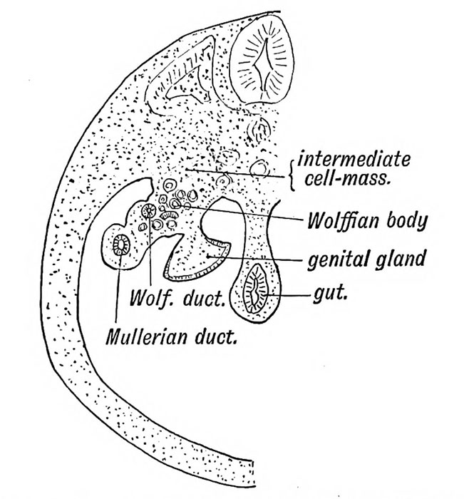
The image shows the positions of the Wolffian and Mullerian ducts in the undifferentiated gonad before the Mullerian duct is degenerated and the Wolffian duct is differentiated.
--[[User:Z8600021|Mark Hill] Your summary is well written, but where is your source citation. In scientific writing you cannot leave out your reference materials, this impacted on your final mark. Historic figure is fine. (3/5)
Lab Assessment 9
Group 1 – Good use of visual aids especially the table as the information is clearly visible, the labelled diagrams are very useful as you can visually see the information that has been written about in the text. Possibly put all of the references at the bottom of the page so they do not interrupt the factual text. The diagram of the schematic of lung disease and the lung models could be incorporated more into relevant text rather than being a stand along diagram so that the diagram can be used to enforce what has been said in the text.
Group 3 - Timeline is very clear and easily shows the order of developmental events that occur. Good detailed information about the development of the foregut, midgut and hindgut however more diagrams could be used in the foregut section. Good description of the deformities that can occur and the possible causes however there is no reference for the possible causes of gastroschisis. The recent findings could do with a little more detail or possibly reference another recent finding.
Group 4 -The table of development is good and visual however to make it look clearer the formatting could be slightly changed so that all of the text is in the same style and format. Nice use of video as well as diagrams. Historic findings could benefit from some diagrams, bullet points or subheadings in order to break up the large amount of text in order to make it easier to read. Good list of all the abnormalities with the relevant information. You could put all of the references at the end to make the main text flow better.
Group 5 – Good use of table and images to map out the stages of skin development. Try and wrap the text around the diagrams to incorporate them more into the text. Good descriptions of abnormalities associated with integumentary development. You could put all of the references at the end to make the main text flow better. The descriptions about development are detailed and easy to understand which is good.
Group 6- Diagrams that have been used are good and show what the text has been explaining. Tables have been used well to illustrate some of the information; tables could also possibly be used to show the developmental timeline more clearly. References could all be collated at the end of the project to make the text flow better from one section to another.
Group 7 – Good use of bullet points and images to clearly show the stages that occur during development. Table is effective at showing the visible anatomical details at different weeks. More work needs to be done on the spinal cord development and meninges development sections. The references in the current research models and findings need to be expanded or put into the references section at the end.
Group 8 – The project is split up into different sections well but you need to include an introduction to your project. Really good information and references but use bullet points and diagrams to break up the text so that it is easier to read. There is good information on DMD but you could possibly write about another abnormality linked to muscle development.
--[[User:Z8600021|Mark Hill] These are good peer assessments, they are though very brief. (8/10)
Lab Assessment 10
<pubmed>24803588</pubmed>
Taste receptor cells detect chemicals that are in the oral cavity and the information is passed to the brain as an output signal. The taste receptors cells mediate the sense of taste and they located in taste buds, each taste bud contains 50-150 taste receptor cells. On the tongue there are distinct areas called papillae where the taste buds are present; there are three different papillae, the fungiform papillae, the circumvallate (CV) papillae and the foliate papillae. This study focuses on the development of the circumvallate (CV) papillae and the factors that control the developmental pathway. It has been found that the development of CV taste placodes is linked to the expression of the transcription factor Wilms' tumor 1 (WT1). Mice with WT1 knockout have defects in the development of CV papillae.
In this experiment WT1-KO mice were used, out of the 263 embryos only 7 survived to birth. qPCR and ChIP experiments were done on taste-enriched tissues from the back half of the tongues at stage E15.5. For the P0 mice taste samples were taken from the epithelium layer of the tongue and these were also analysed. Taste buds from adult mice were also analysed.
The study found that WT1 is needed in order for the normal expression of Ptch1and Lef1 for CV papillae development. It was also found that SOX2 expression is disturbed when there is an absence of WT1; SOX2 is needed for the development of the placode in the fungiform papillae. WT1 is expressed in adult taste buds which suggest that WT1 is needed for the formation and maintenance of taste cells. In addition WT1 is also need to regulate the expression of BMP4 in embryonic taste cells.
https://embryology.med.unsw.edu.au/embryology/index.php/Sensory_-_Taste_Development
--[[User:Z8600021|Mark Hill] This is a very good recent paper. (5/5)

