User:Z5018221
| Student Information (expand to read) | ||||||||||||||||||||||||||||||||||||||||||||||||||||||||||||||||
|---|---|---|---|---|---|---|---|---|---|---|---|---|---|---|---|---|---|---|---|---|---|---|---|---|---|---|---|---|---|---|---|---|---|---|---|---|---|---|---|---|---|---|---|---|---|---|---|---|---|---|---|---|---|---|---|---|---|---|---|---|---|---|---|---|
| Individual Assessments | ||||||||||||||||||||||||||||||||||||||||||||||||||||||||||||||||
|
Please leave this template on top of your student page as I will add your assessment items here. Beginning your online work - Working Online in this course
Click here to email Dr Mark Hill | ||||||||||||||||||||||||||||||||||||||||||||||||||||||||||||||||
| Lab 1 Assessment - Researching a Topic | ||||||||||||||||||||||||||||||||||||||||||||||||||||||||||||||||
In the lab I showed you how to find the PubMed reference database and search it using a topic word. Lab 1 assessment will be for you to use this to find a research reference on "fertilization" and write a brief summary of the main finding of the paper.
| ||||||||||||||||||||||||||||||||||||||||||||||||||||||||||||||||
| Lab 2 Assessment - Uploading an Image | ||||||||||||||||||||||||||||||||||||||||||||||||||||||||||||||||
OK you are now in a group
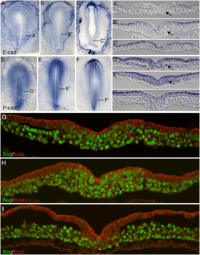
Initially the topic can be as specific or as broad as you want. Chicken embryo E-cad and P-cad gastrulation[1] References
| ||||||||||||||||||||||||||||||||||||||||||||||||||||||||||||||||
| Lab 4 Assessment - GIT Quiz | ||||||||||||||||||||||||||||||||||||||||||||||||||||||||||||||||
|
ANAT2341 Quiz Example | Category:Quiz | ANAT2341 Student 2015 Quiz Questions | Design 4 quiz questions based upon gastrointestinal tract. Add the quiz to your own page under Lab 4 assessment and provide a sub-sub-heading on the topic of the quiz. An example is shown below (open this page in view code or edit mode). Note that it is not just how you ask the question, but also how you explain the correct answer. | ||||||||||||||||||||||||||||||||||||||||||||||||||||||||||||||||
| Lab 5 Assessment - Course Review | ||||||||||||||||||||||||||||||||||||||||||||||||||||||||||||||||
| Complete the course review questionnaire and add the fact you have completed to your student page. | ||||||||||||||||||||||||||||||||||||||||||||||||||||||||||||||||
| Lab 6 Assessment - Cleft Lip and Palate | ||||||||||||||||||||||||||||||||||||||||||||||||||||||||||||||||
| ||||||||||||||||||||||||||||||||||||||||||||||||||||||||||||||||
| Lab 7 Assessment - Muscular Dystrophy | ||||||||||||||||||||||||||||||||||||||||||||||||||||||||||||||||
| ||||||||||||||||||||||||||||||||||||||||||||||||||||||||||||||||
| Lab 8 Assessment - Quiz | ||||||||||||||||||||||||||||||||||||||||||||||||||||||||||||||||
| A brief quiz was held in the practical class on urogenital development. | ||||||||||||||||||||||||||||||||||||||||||||||||||||||||||||||||
| Lab 9 Assessment - Peer Assessment | ||||||||||||||||||||||||||||||||||||||||||||||||||||||||||||||||
| ||||||||||||||||||||||||||||||||||||||||||||||||||||||||||||||||
| Lab 10 Assessment - Stem Cells | ||||||||||||||||||||||||||||||||||||||||||||||||||||||||||||||||
As part of the assessment for this course, you will give a 15 minutes journal club presentation in Lab 10. For this you will in your current student group discuss a recent (published after 2011) original research article (not a review!) on stem cell biology or technology.
| ||||||||||||||||||||||||||||||||||||||||||||||||||||||||||||||||
| Lab 11 Assessment - Heart Development | ||||||||||||||||||||||||||||||||||||||||||||||||||||||||||||||||
| Read the following recent review article on heart repair and from the reference list identify a cited research article and write a brief summary of the paper's main findings. Then describe how the original research result was used in the review article.
<pubmed>26932668</pubmed>Development | ||||||||||||||||||||||||||||||||||||||||||||||||||||||||||||||||
| ||||||||||||||||||||||||||||||||||||||||||||||||||||||||||||||||
Lab Attendance
Z5018221 (talk) 14:41, 12 August 2016 (AEST)
Z5018221 (talk) 14:06, 19 August 2016 (AEST)
Z5018221 (talk) 14:13, 26 August 2016 (AEST)
Z5018221 (talk) 15:19, 2 September 2016 (AEST)
Z5018221 (talk) 13:21, 9 September 2016 (AEST)
Z5018221 (talk) 13:05, 16 September 2016 (AEST)
Z5018221 (talk) 14:43, 23 September 2016 (AEST)
Z5018221 (talk) 13:54, 7 October 2016 (AEDT)
Lab 1 Assessment
<pubmed>27462598</pubmed>
During the process of oocyte retrieval, a technique used in in vitro fertilization (IVF), oocytes may become exposed to ovarian endometriotic fluid, an event that is unlikely however possible in the clinical environment. Hashin et al. investigated the effect of various concentrations of human endometriotic fluid exposure on mice cumulus-oocyte complexes (COCs) by measuring fertilization, blastocyst formation and hatched blastocyst rate. COCs obtained from 46-week-old female mice were randomly divided into 5 groups and each exposed to differing concentrations of endometriotic fluid obtained from a 29-year-old patient during aspiration, for 5 minutes. Final concentrations were 0.625%, 1.25%, 2.5% and 5%. COCs were then inseminated with sperm retrieved from male mice. Fertilisation was assessed day 1 following insemination by formation of 2-cell, and blastocyst formation on day 5.
Results indicated no apparent difference between the endometriotic fluid treated groups and the non-exposed control group except for the 0.625% exposure group which had a higher fertilization rate. Blastocyst formation rate and hatched blastocyst rates were also similar between exposed and non-exposed groups, overall indicating no detrimental impact on fertilization, blastocyst formation, and hatched blastocyst rates, following exposure to differing concentrations of endometriotic fluid. Previous experiments have generated results proving otherwise such as an experiment by Piromlertamorn et al. using mice. It was shown that use of a single concentration of endometriotic fluid leads to similar results in fertilization and blastocyst formation rate, however a significant decrease in blastocyst hatching rate. Despite the result, the authors noted this decrease was also observed in serum-treated groups, providing conflicting results. Hashin et al. have additionally noted a few limitations to their experiment where the endometriotic fluid utilised was only from a single patient, and pregnancy and implantation rates were not analysed. The authors of the study stress that to truly assess the effect of endometriotic fluid on oocyte and embryonic development, the contents of the fluid should be studied further to determine cytoxic effect, and its effect on pregnancy and implantation.
| Mark Hill 18 August 2016 - You have added the citation correctly and written a good descriptive summary of the article findings. I guess I am am wondering why the researchers expected human endometriotic fluid to have an effect on mice? Note the house mouse generally only lives about a year. | Assessment 5/5 |
Lab 2 Assessment
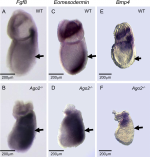
Ago2 and it's Importance in FGF Signalling in Mammalian Gastrulation
<pubmed>18166081</pubmed>
| Mark Hill 29 August 2016 - All information Reference, Copyright and Student Image template correctly included with the file and referenced on your page here. Note though to display the reference citation correctly with the legend. You need to include the ref name for a citation, as shown below.
Code: <ref name="PMID18166081"><pubmed>18166081</pubmed></ref> Ago2 in Mammalian Gastrulation[1] |
Assessment 5/5 |
Referencing
Z5018221 (talk) 14:34, 5 August 2016 (AEST)
PMID 27486480
http://molecularcytogenetics.biomedcentral.com/articles/10.1186/s13039-016-0269-1
Lab 3 Assessment
| Mark Hill 31 August 2016 - Lab 3 Assessment Quiz - Mesoderm and Ectoderm development. | Assessment 3/5 |
Lab 4 Assessment
Gastrointestinal Development in the Embryo
| Mark Hill 14 September 2016 - These seem good quiz questions. Note that question 1 your answer says "The yolk sac provides nutrients and functions as the circulatory system for the early embryo.", the yolk sac does not really provide nutrients in placental animals, only in birds and reptiles, it does though contribute to cardiovascular development. | Assessment 5/5 |
Lab 5 Assessment
Questionnaire completed
| Mark Hill 22 September 2016 - Questionnaire on course structure. | Assessment 5/5 |
Lab 6 Assessment
Cleft palate, often occurring in conjunction with cleft lip is among the most common congenital malformations and involves abnormal facial development during gestation. Between the 4th week and the 6th-7th weeks of development, migrating neural crest cells of the anterior neural tube with the mesodermal cells form the facial primordia. From here the filtrum and primary palate are formed, by the merging of the nasal prominences to form the intermaxillary segment, which then fuses with the maxillary prominences to form the upper lip. The palatal shelves additionally grow out from the maxillary prominences.
This complex process involves a variety of signalling molecules, transcriptions factors and cell-cell interactions, and an interference in the cascade can lead to the failure of fusion of the facial primordia, resulting in facial clefting. An important transcription factor expressed primarily in the palatal shelves and tongue during palatogenesis is TBX22. TBX22 is a member of a family of transcriptional regulators containing a common DNA-binding domain, the T-box. Mutations of the TBX22 gene have been strongly linked with syndromic X linked cleft palate producing clefting of the palate and ankyloglossia, otherwise known as tongue-tie.
Mutations of the TBX22 gene commonly occur as nucleotide alterations where amino acid substitutions, deletions and transitions may occur as well as sequence variants close to splice sites. These mutations lead to formation of truncated proteins, with missense mutations affecting amino acids within the DNA binding T box domain. Missense mutations of TBX22 primarily occur at major areas of contact with the target DNA, affecting and reducing the protein’s ability to bind to the DNA or even other binding proteins in the transcriptional complex. This loss in binding to DNA and other crucial proteins within the transcriptional complex therefore affects the downstream cascade of gene activation, and as a result the development and fusion of the palate.
Through research it has been shown that a variety of genetic mutations involving genes such as TBX22, PVRL1, and IRF6 lead to facial clefting. However the location and type of mutation does not completely determine the severity of the clefting but rather environmental factors. Environmental factors such as alcohol, tobacco, and toxin exposure during pregnancy can also impact palatogenesis.
1) <pubmed>14729838</pubmed> 2) <pubmed>14722155</pubmed>
| Mark Hill 13 October 2016 - TBX genes act as transcription factors regulating many developmental patterning processes throughout the embryo.
OMIM TBX22 has been identified as expressed in the palatal shelves during the elevation process. |
Assessment 5/5 |
Lab 7 Assessment
1) What is/are the dystrophin mutation(s)?
In approximately 65% of patients with Duchenne Muscular Dystrophy (DMD), lack of dystrophin is due to a frameshift mutation caused by gene deletions, which give rise to premature stop codons, hence preventing translation to protein. In the remaining cases, point mutations and small gene insertions/deletions are the underlying causes leading to formation of truncated protein, with exon duplications responsible for approximately 5% of all mutations.
2) What is the function of dystrophin?
DMD is caused by the absence of the protein dystrophin in skeletal, smooth and cardiac muscle, a protein present at the membrane of muscle fibres and an important link between the cytoplasmic and extracellular matrix of the muscle fibre. Due to this it has an important mechanical role where it maintains the strength of muscle fibres, as, in fibres lacking dystrophin, normal contraction weakens the fibre and leads to degeneration. As well as playing a mechanical role, it is also involved in signalling pathways such as pathways for production of nitric oxide, and the Ras/MPAK pathway.
3) What other tissues/organs are affected by this disorder?
Dystrophin is not only present in skeletal muscle, but also in cardiac muscle. The heart can be affected whereby the conduction tissues can get damaged and lead to abnormal rhythms such as arrthymia and dysrhythmia, or pump abnormally leading to cardiomyopathy. Weakening of respiratory muscles is additionally seen in patients, leading to difficulties in breathing. At this point ventilation machines are utilized to assist with breathing.
4) What therapies exist for DMD?
Over the years, patients with DMD have begun to live longer into their 20s and 30s showing an improvement in treatment. Various factors such as earlier and continuous use of corticosteroids, routine flu and pneumococcal injections, and intense physical therapy have influenced these results. Currently, the therapies available for DMD aim to manage the disease rather than cure it. Corticosteroids are given to the patient to improve muscle function and strength however with continued use into adulthood, side effects such as weight gain, hypertension, hyperglycemia, cataracts and osteoporosis begin to surface. Patients also undergo physiotherapy to maintain function of the limbs, particularly of the upper extremity. A daily stretch routine targeting the muscles of the forearm wrist and fingers is crucial so the patient can still control their wheelchair, for example.
Research continues in the field of the disease and potential therapies have been proposed, in particular gene therapies. An example of a potential gene therapy for DMD patients is the CRISPR/Cas 9 system. This system is a genome modification technology, first identified in bacteria, re-engineered to 2 components of an RNA guide sequence and a DNA endonuclease. The system has now been modified to function in mammalian cells where it could generate random mutations or targeted repair, hence has been proposed as a potential way of disease modification.
In order to correct the mutant phenotype of DMD the CRISPR/Cas9 system was recently employed on a mouse MdX model. It was found that the CRISPR/Cas 9 was able to repair a large population of skeletal myocytes, with the myocytes showing a good survival rate. Although the CRISPR/Cas 9 system has been successful in animal studies, further research must now be undertaken for employment in human patients.
5) What animal models are available for muscular dystrophy?
Currently, the most common animal model used for DMD research is the MDX mouse, which contains a nonsense mutation abolishing formation of dystrophin. However to bridge the gap between these models and human patients in terms of vector production and immune response, canine models are now being utilized. Dog breeds of the Golden Retriever, Rottweiler and Cavalier King Charles have been reported as dystrophin deficient due to gene deletions.
References
1) <pubmed>27515321</pubmed> 2) <pubmed>16887341</pubmed> 3) <pubmed>27619714 </pubmed>
| Mark Hill 13 October 2016 - Comprehensive discussion of Muscular Dystrophy. I liked that you included the CRISPR/Cas 9 application. You could have included the specific mutations in the animal models. | Assessment 5/5 |
Lab 9 Assessment
These are good reviews of the project pages, with some specific examples. They include a balanced critical assessment, given the existing status of these pages. 8/10
Team 1
In terms of the topic of WnT signalling pathway, the page is beginning to come together with a great amount of information. What I particularly like is how the different concepts introduced in the page have been explained for e.g. the different WnT pathways. However the content for each pathway does not seem to be consistent. While the canonical pathway addresses the mechanism, the non-canonical one doesn’t. I would suggest constructing a table to compare the similarities and differences between the various pathways, and adding images or shorts clips with audio to represent the elements of the pathways in a different form. This would not only enhance the look of your page but also make it more interactive for the audience.
A great positive is to see links to research articles have been provided for the audience to access if they are interested to read on further. The links are short and easy to see, and direct you straight to the article on Pubmed, a reliable source. An effort has also been made to summarise the article, however the summary should be available as a simple breakdown so the audience can refer to it if they struggle to understand. The summaries include some jargon that can be further simplified.
In relation to criteria 1, the key points have definitely been highlighted and the signalling pathway has been associated with the fetus, however to make it more interesting and satisfy criteria 5, possibly construct a table or briefly outline how WnT signalling is involved in other areas such as Type 2 Diabetes and Cancer. Furthermore, to relate the topic back to embryological development explore the pathway in areas such as gastrulation rather than only talking about skin development.
Lastly to satisfy criteria 3, attempt to include in text citations within the paragraphs, instead of displaying references towards the end of the page. Overall, great job in gathering and highlighting key features, and backing up your information with relevant articles!
Team 2
A job well done with the introduction! The introduction is brief however manages to link the topic to embryonic development, different medical conditions, whilst also outlining the function and elements of the pathway. Being brief and succinct, it allows the viewer to continue exploring the page without experiencing confusion at the first lot of information. Further to this, the history of the pathway is formatted well and is not too overwhelming or boring. It is evident you have decided ‘Current Areas of Research’ will also be included in your page which is a great idea as you have included a section on History. This would ensure your Wiki flows well, and covers the pathway from start to finish.
Images have been included to visually represent the elements of the general pathway, as well as the elements specific to the pathway in cardiac development, which forms a great aid for viewers in understanding the content. Videos explaining the different canonical and non- canonical pathways could also be included for viewers with a video learning preference.
Use of in text citations neatens the layout of information and enables viewers to access the article should they find the point interesting. Numerous subheadings have been included which further break down the page into small sections of information. This is a fantastic positive as viewers can locate information in which they are interested in easily instead of having to read through long paragraphs of text. I feel as if linking the topic clinically is extremely important which you have done a great job in! Along with the text explaining the disease, you could possibly include a table stating the disease, the mutation, and the symptoms for viewers after a more easy, accessible format.
Throughout the topics covered, a lot of jargon is utilized, however a full glossary has not been provided. A glossary should definitely be included for terms such as ‘gastrulation’, ‘kinases’ or ‘cardiogenesis’ in order to satisfy criteria 4. Additionally, the page does not cater for viewers interested in further reading up on the topic. To ensure criteria’s 4 and 5 are met, links to interesting facts or articles could be provided so the audience has access to more information if they would like to further their understanding. With a few improvements this Wiki page can definitely prove helpful in understanding the pathway!
Team 3
A great introduction to the topic, allowing the reader to slowly transition into the more in-depth points! I particularly like how you have broken down the different constituents of the pathway such as the receptors and protein subtypes and provided a succinct table outlining their function and clinical significance before moving onto the mechanism. Although the ‘FGF Subtype’ table has proven to be effective and helpful, the table on ‘History’ does not seem to be thorough and is very limited. Possibly extending the table by researching more developments in the field of FGF Signalling could make it appear more complete.
Effort has been made to include a hand drawn image of the signalling pathway, which serves as a great source of aid in understanding how the pathway works whilst reading the text beside it. In saying that, effort should be further made to include a complete glossary and ensure terms such as ‘receptor dimerization’ ‘morphogenesis’ are broken down for the reader in order to satisfy criteria 4. This is not only seen in the ‘Signal Transduction’ section but also throughout the other sections. As you have included a fantastic image on bone development to represent the information visually, it would also be a good idea to post up images covering the other areas of embryonic development, such as kidney and inner ear development! You could even consider including short clips explaining these processes to make the page more interactive.
It is clear a decision has been made to talk about ‘Animal Models’. As well as including text on the topic, a possible option could be including a table briefly outlining which animal model has contributed to what knowledge in relation to the pathway in order to simplify the information.
A particular highlight of the Wiki page is the use of a quiz. It is great to see viewers can test their understanding of the topic towards the end and challenge themselves! For the correct option to each question a link to a supporting article or particular section of the page can be provided so the viewer can revisit the information should they have answered the question incorrectly. Overall a great use of tables, images and interactive components!
Team 5
Well done on constructing a thorough Wiki page on the topic of T-box Genes and their Signalling! Viewing the page it is evident numerous headings and subheadings have been provided to accommodate for the large amount of information gathered. Starting off with the introduction I like how you have included a section on what T’-box exactly means, however the information provided in this section talks about the history significantly, hence to turn this into a positive I would suggest adding a table or timeline outlining the major events and discoveries in the past to present this information in a complete, meaningful way. This issue is also seen with the section ‘Origins of the T-box genes’ where major discoveries are highlighted and in which year they occurred. This information can also merge with the history timeline/table.
Within the ‘What does T-box mean?’ section you have also added information on which animal studies were undertaken for the discoveries. To avoid spreading of information and causing confusion for the reader, you could either construct a table to show which animal study was completed in which year, and what discovery it led to as 3 columns, or bring this information down to the section ‘Animal Models’. In saying that, you have attempted to utilize a table and the table works very well with the topic of the different T-box genes, and would prove great help for the viewer.
It is great to see T-box genes and Signalling has been explored further in the field of embryonic development. Extensive information is provided with good use of in- text citations, allowing the reader to navigate to relevant articles. The ability to navigate could be further improved by providing an accessible link to the ‘Abnormalities’ section in a case where you are directing the reader to the section for further information, instead of plain text. Beneath each section for e.g. ‘Limb Development’ Pubmed links have been provided to relevant articles, which is a fantastic idea, however the links have no indication whatsoever of what the article is about. You could add a sentence each next to the links briefly stating what the article is exploring in relation to limb development.
Overall with a few more images, possibly some interactive components such as clips, and a knowledge testing short exam or quiz this Wiki page will stand out. Remember to ensure your information flows well by placing it within appropriate sections!
Team 6
It is great to see an in-depth overview of the TGF beta Signalling pathway and it’s mechanism of action. The page is off to a great start and with a few key improvements it can turn into a successful one! Firstly, I like the use of images to aid the reader in understanding the content better should they be a visual learner. By reading the text on the way the pathway works it is clear it is a complicated process hence a suggestion would be to add short clips/animations to simplify it for the viewer.
Under your introduction an attempt has been made to briefly highlight the main features of the pathway, however a negative of this section is that a lot of the content is basically listed. For example it is mentioned TGF-beta is part of a larger TGF superfamily comprising of different members such as activins and GDFs. Instead of listing, try presenting your information in a different format such as a table with s few columns stating the member, it’s function, and possibly what a mutation could lead to.
Additionally it is also stated that TGF beta has certain functions such as controlling angiogenesis however doesn’t expand on the ‘other’ functions it has. To turn this into a positive, dedicate different sections on how the pathway is involved in angiogenesis, hormone secretion, proliferation etc. If there are too many functions to fit onto the page, you could shift a few to your ‘Further Reading’ section as an option if the reader would like to explore further into the pathway’s functions, or construct a hidden table that the viewer can expand if they wish to.
In order to satisfy criteria 3, in text citations should be incorporated within text so the viewer has an option to access the article if they find the statement interesting. A reference list has been posted with a few references however ensure they are cited in the correct format. Overall a good start!
| Lab 10 - Stem Cell Presentations 2016 | |
|---|---|
| Group Mark | Assessor General Comments |
|
Group 1: 15/20 Group 2: 19/20 Group 3: 20/20 Group 4: 19/20 Group 5: 16/20 Group 6: 16/20 |
The students put great effort in their presentation and we heard a nice variety of studies in stem cell biology and regenerative medicine today. The interaction after the presentation was great.
As general feedback I would like to advise students to:
|
Lab 11
0/5
Heart development paper?
- ↑ <pubmed>18166081</pubmed>


