Talk:Carnegie stage 13
Carnegie Collection
1 19 87 76 112 116 148 186 239 248 407 463 523 588 786 800 808 826 836 963 1075 3956 4046 5541 5682 5874 6032 6469 6473 7433 7618 7669 7889 8066 8119 8147 8239 8372 8581 8967 9296 9297 9697
Image Galleries
Bright Field


Scanning EM
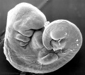
- Stage13 sem4.jpg
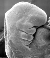
- Stage13 sem5.jpg
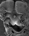
- Stage13 sem6.jpg


3D Animation
- 6mm Pig Embryo - approximately Human day 32 (Carnegie Stage 13/14 embryo) Stage13_3d01.flv
2004
A social biography of Carnegie embryo no. 836
Anat Rec B New Anat. 2004 Jan;276(1):3-7.
Morgan LM. Department of Sociology and Anthropology, Mount Holyoke College, 50 College Street, South Hadley, MA 01075-1426, USA. lmmorgan@mtholyoke.edu
Abstract
A tiny, sectioned embryo specimen known as Carnegie no. 836 has served as the prototype for Stage 13 (28-32 days) since the 1910s. Recently digitalized and reanimated for the 21st century, this singular specimen is now being used to develop 3D and 4D visualizations. Yet the social origins of the specimen have been largely forgotten. This essay traces the biography of 836 from its origins in a young woman's life, through sectioning and transformation into a scientific specimen, to its contemporary manifestations as a symbol of life. By reuniting the specimen with its story, we can appreciate how cultural attitudes toward embryo specimens have changed over the past century.
Copyright 2004 Wiley-Liss, Inc.
PMID 14750188
The development of the human brain from a closed neural tube at stage 13
Anat Embryol (Berl). 1988;177(3):203-24.
Müller F, O'Rahilly R. Source Carnegie Laboratories of Embryology, California Primate Research Center, Davis 95616.
Abstract
Twenty-five embryos of stage 13 (28 days) were studied in detail and graphic reconstructions of seven of them were prepared. Thirty or more somitic pairs are present, and the maximum is possibly 39. The notochord is almost entirely separated from the neural tube and the alimentary epithelium, and its rostral tip is closely related to the adenohypophysial pocket. Caudal to the cloacal membrane, the caudal eminence is the site of secondary neurulation. The eminence, which usually contains isolated somites, is the area where new notochord, hindgut, and neural tube are forming. The neural cord develops into neural tube without the intermediate phase of a neural plate (secondary neurulation). Canalization is regular and the lumen is continuous with the central canal. The neural tube is now a closed system, filled with what may be termed "ependymal fluid." The brain is widening in a dorsoventral direction. Neuromeres are still detectable. The following features are distinguishable: infundibular area of D2, chiasmatic plate of D1, "adult" lamina terminalis, and commissural plate (at levels of nasal plates). The beginning of the synencephalon of D2 can be discerned. The retinal and lens discs are being defined. The mesencephalic flexure continues to diminish. The midbrain possesses a sulcus limitans, and the tegmentum may show the medial longitudinal fasciculus. The isthmic segment is clearly separated from rhombomere 1. Lateral and ventral longitudinal fasciculi are usually present in the hindbrain, and the common afferent tract is beginning. Somatic and visceral efferent fibres are seen in certain nerves: 6, 12; 5, 7, 9-11. The first indication of the cerebellum may be visible in the alar lamina of rhombomere 1. The terminal-vomeronasal crest appears. Various cranial ganglia (e.g., vestibular, superior ganglia of 9, 10) are forming. The trigeminal ganglion may show its three major divisions. Epipharyngeal placodes of pharyngeal arches 2 to 5 contribute to cranial ganglia 7, 9, and 10. The spinal neural crest is becoming segregated, and the spinal ganglia are in series with the somites. Ventral spinal roots are beginning to develop.
PMID 3354839