Uploads by Z5093005
From Embryology
This special page shows all uploaded files.
| Date | Name | Thumbnail | Size | Description | Versions |
|---|---|---|---|---|---|
| 12:52, 26 October 2017 | SchizencephalicB.jpg (file) |  |
1.37 MB | ===Description:=== These features are observable in brain scans of patients with Microcephaly. ===Copyright:=== Limited License to use press release material according to: http://service.prweb.com/legal/copyright/ {{Template:Student Image}} ===Refere... | 1 |
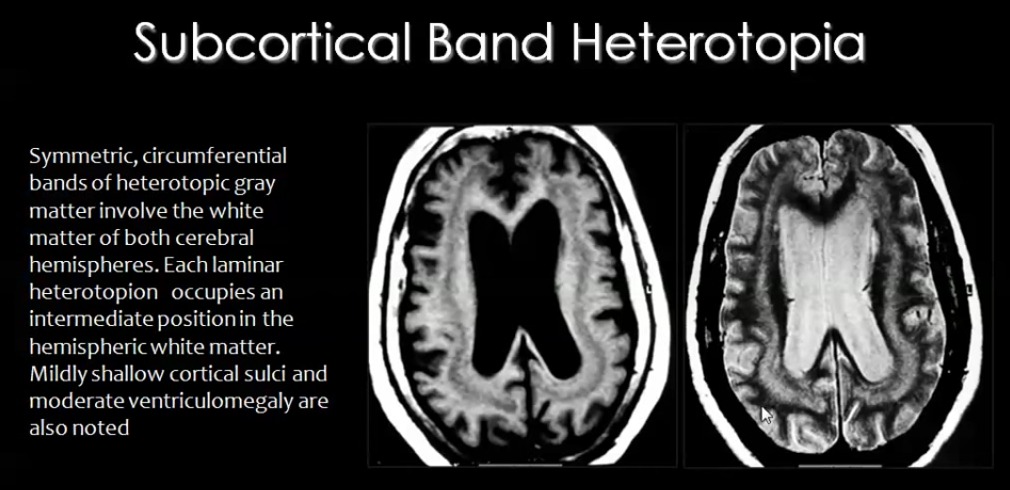
| 12:47, 26 October 2017 | Heteroptopia.png (file) |  |
451 KB | ===Description:=== Sub-cortical Band Heterotopia- abnormal locations of neurons form nodules or bands in the white matter of both cerebral hemispheres. ===Copyright:=== Creative Commons License {{Template:Student Image}} ===Reference: === <ref> Mahfo... | 1 |
| 12:44, 26 October 2017 | Image.png (file) |  |
129 KB | ===Description:=== These features may be observable occasionally in patients with Microcephaly. ===Copyright:=== Creative Commons Attribution {{Template:Student Image}} ===Reference: === <ref>USDA & Felipe Dana/AP and Creative Commons License(CC) (20... | 1 |
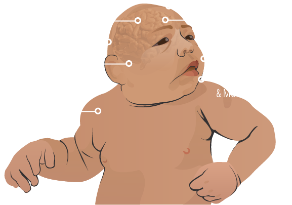
| 12:43, 26 October 2017 | Symptoms of microcephaly.png (file) |  |
129 KB | ===Description:=== These features may be observable occasionally in patients with Microcephaly. ===Copyright:=== Creative Commons Attribution {{Template:Student Image}} ===Reference: === <ref>USDA & Felipe Dana/AP and Creative Commons License(CC) (20... | 2 |
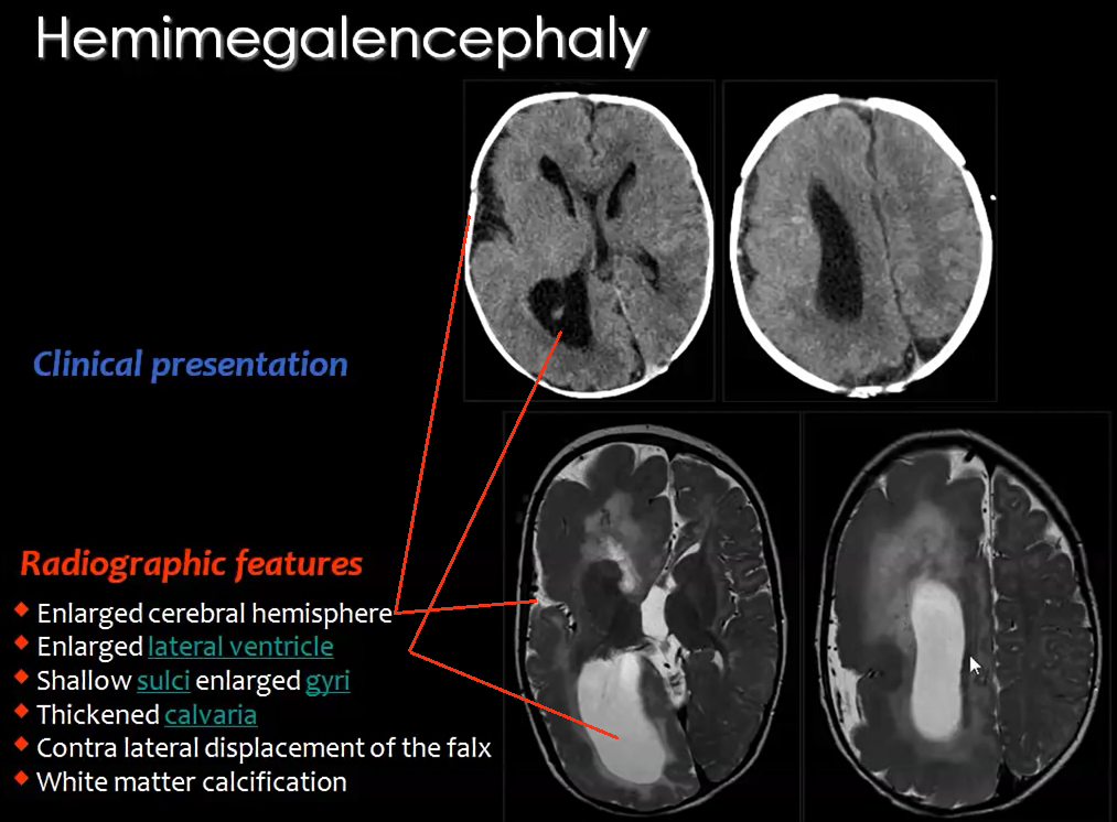
| 12:39, 26 October 2017 | Hemimegalencephaly2.png (file) |  |
510 KB | ===Description=== Radiographic features seen in Hemimgalencephaly. ===Copyright=== Creative Commons License {{Template:Student Image}} ===Reference: === <ref>Mahfouz, M. (2015). Imaging of cortical formation disorders - DRE 4 - Prof. Dr Mamdouh Mahf... | 1 |
| 16:17, 24 October 2017 | Brain sectin showing cortex.jpg (file) |  |
25 KB | ===Description:=== Section of human brain- anatomical sections. The cerebral cortex is gray, this is why the brain appears gray. ===Copyright:=== Creative Commons Attribution Share-Alike 3.0 License. ===Reference:=== <ref>Mikayla D (2017). 23. [image... | 1 |
| 15:30, 24 October 2017 | Intro section cortex.jpg (file) |  |
111 KB | ===Description=== The cerebral cortex is the outermost layer of the cerebral hemispheres. It appears gray and contains cell bodies of neurons. ===Copyright:=== Allowed to use non-commercially according to http://slideplayer.com/support/terms/ "Exce... | 1 |
| 15:27, 24 October 2017 | Introcortex.jpg (file) |  |
111 KB | ===Description=== The cerebral cortex is the outermost layer of the cerebral hemispheres. It appears gray and contains cell bodies of neurons. ===Copyright:=== Allowed to use non-commercially according to http://slideplayer.com/support/terms/ "Except... | 3 |
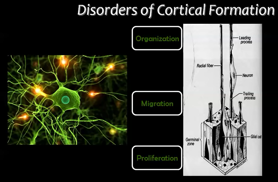
| 19:48, 23 October 2017 | Stages of development.png (file) |  |
495 KB | ====Description==== This image shows the those layers of the cerebral cortex which correspond to the main stages of cortical development. These stages of development are where abnormalities may arise in the developing fetus. ====Copyright==== Creative... | 1 |
| 19:44, 23 October 2017 | Disorders of Cortical Formation2.png (file) |  |
495 KB | ====Description==== This image shows the those layers of the cerebral cortex which correspond to the main stages of cortical development. These stages of development are where abnormalities may arise in the developing fetus. ====Copyright: ==== Creati... | 2 |
| 17:52, 23 October 2017 | Cortex-group project.jpg (file) |  |
25 KB | '''Description:''' Section of human brain- anatomical sections. The cerebral cortex is gray, this is why the brain appears gray. '''Copyright:''' Creative Commons Attribution Share-Alike 3.0 License. '''Reference:''' <ref> Mikayla D (2017). 23. [imag... | 1 |
| 17:04, 23 October 2017 | Intro cortex.jpg (file) |  |
111 KB | The cerebral cortex is the outermost layer of the cerebral hemispheres. It appears gray and contains cell bodies of neurons. Copyright: Allowed to use non-commercially according to http://slideplayer.com/support/terms/ "Except as otherwise provided, t... | 1 |
| 18:40, 5 October 2017 | SchizencephalicBrain.jpg (file) |  |
1.37 MB | Schizencephaly Copyright: Limited License to use press release material according to: http://service.prweb.com/legal/copyright/ Reference: <ref>PRWeb (2013). Schizencephalic Brain. [image] Available at: http://www.prweb.com/releases/2013/7/prweb3350... | 1 |
| 17:30, 5 October 2017 | SBH.png (file) |  |
451 KB | Sub-cortical Band Heterotopia- abnormal locations of neurons form nodules or bands in the white matter of both cerebral hemispheres Copyright: Creative Commons License Reference: <ref>Mahfouz, M. (2015). Imaging of cortical formation disorders - DRE... | 1 |
| 16:49, 5 October 2017 | Symptoms-microcephaly.png (file) |  |
66 KB | Symptoms seen in Microcephaly Copyright: Creative Commons Attribution Reference: <ref>USDA & Felipe Dana/AP and Creative Commons License(CC) (2017). Understanding Zika. [online] Goinvo.com. Available at: http://www.goinvo.com/features/zika/ [Accesse... | 1 |
| 16:40, 5 October 2017 | Hemimegalencephaly.png (file) |  |
510 KB | Hemimegalencephaly Copyright: Creative Commons License Reference: <ref>Mahfouz, M. (2015). Imaging of cortical formation disorders - DRE 4 - Prof. Dr Mamdouh Mahfouz. [video] Available at: https://www.youtube.com/watch?v=l_nTggR7LTE [Accessed 23 Sep... | 1 |
| 15:55, 5 October 2017 | Disorders of Cortical Formation.png (file) |  |
495 KB | Main stages of cortical development where abnormalities may arise "Reference" <br/> <ref>Mahfouz, M. (2015). Imaging of cortical formation disorders - DRE 4 - Prof. Dr Mamdouh Mahfouz. [video] Available at: https://www.youtube.com/watch?v=l_nTggR7LTE [... | 1 |