Uploads by Z5059996
From Embryology
This special page shows all uploaded files.
| Date | Name | Thumbnail | Size | Description | Versions |
|---|---|---|---|---|---|
| 16:27, 25 October 2017 | Development of the Semilunar Valves.jpg (file) |  |
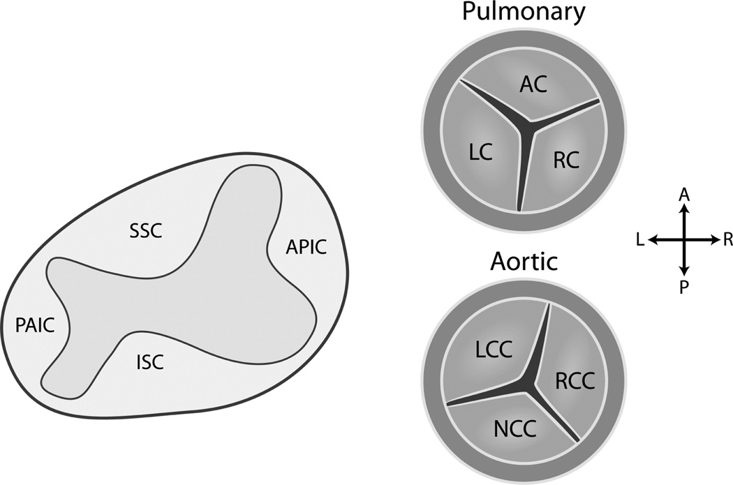
105 KB | Description: Development of the leaflets of the aortic and pulmonary valves. The semilunar valves arise from the conotruncal and intercalated cushions of the outflow tract. The conotruncal (superior and inferior septal) cushions give rise to the right... | 1 |
| 10:43, 25 October 2017 | Hypoplastic Left Heart Syndrome (HLHS).png (file) | .png) |
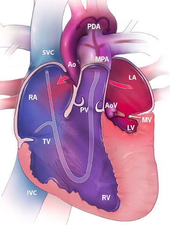
254 KB | Description: Hypoplastic Left Heart Syndrome (HLHS).RA. Right Atrium. RV. Right Ventricle. LA. Left Atrium. LV. Left Ventricle. SVC. Superior Vena Cava. IVC. Inferior Vena Cava. MPA. Main Pulmonary Artery. Ao. Aorta. PDA. Patent Ductus Arteriosus. TV.... | 1 |
| 10:29, 24 October 2017 | Tetralogy of Fallot (TOF).png (file) | .png) |
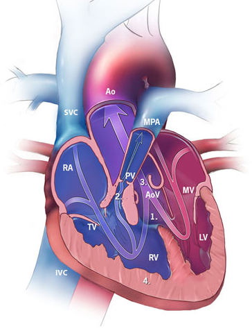
238 KB | Description: Tetralogy of Fallot (TOF). RA. Right Atrium. RV. Right Ventricle. LA. Left Atrium. LV. Left Ventricle. SVC. Superior Vena Cava. IVC. Inferior Vena Cava. MPA. Main Pulmonary Artery. Ao. Aorta. TV. Tricuspid Valve. MV. Mitral Valve. PV. Pulm... | 1 |
| 09:18, 22 October 2017 | Truncus Arteriosus.png (file) |  |
314 KB | Description Truncus Arteriosus. RA. Right Atrium. RV. Right Ventricle. LA. Left Atrium. LV. Left Ventricle. SVC. Superior Vena Cava. IVC. Inferior Vena Cava. MPA. Main Pulmonary Artery. Ao. Aorta. TV. Tricuspid Valve. MV. Mitral Valve Reference http... | 1 |
| 16:01, 21 October 2017 | Atrial Septal Defect (ASD).png (file) | .png) |
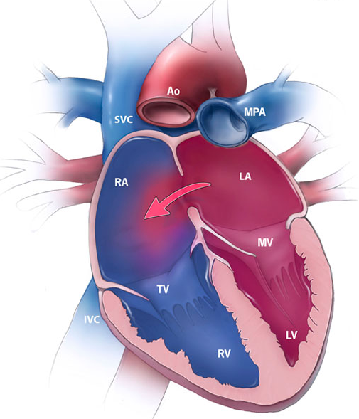
337 KB | Description: Atrial Septal Defect (ASD). RA. Right Atrium. RV. Right Ventricle. LA. Left Atrium. LV. Left Ventricle. SVC. Superior Vena Cava. IVC. Inferior Vena Cava. MPA. Main Pulmonary Artery. Ao. Aorta. TV. Tricuspid Valve. MV. Mitral Valve Refere... | 1 |
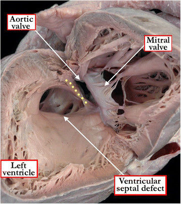
| 13:43, 21 October 2017 | Ventricular Septal Defect (VSD).jpeg (file) | .jpeg) |
46 KB | The view from the left ventricle in this specimen shows a ventricular septal defect with exclusively muscular borders opening towards the outlet of the right ventricle, but with postero-caudal deviation of the muscular outlet septum (yellow dots), caus... | 1 |
| 18:49, 4 October 2017 | The Process of Atrial Septation.png (file) |  |
1.24 MB | Atrial septation. This cartoon depicts the developmental events responsible for the formation of the atrial septal complex (see text). The yellow box in the back of the heart represents the posterior Second Heart Field giving rise to the DMP which in t... | 1 |
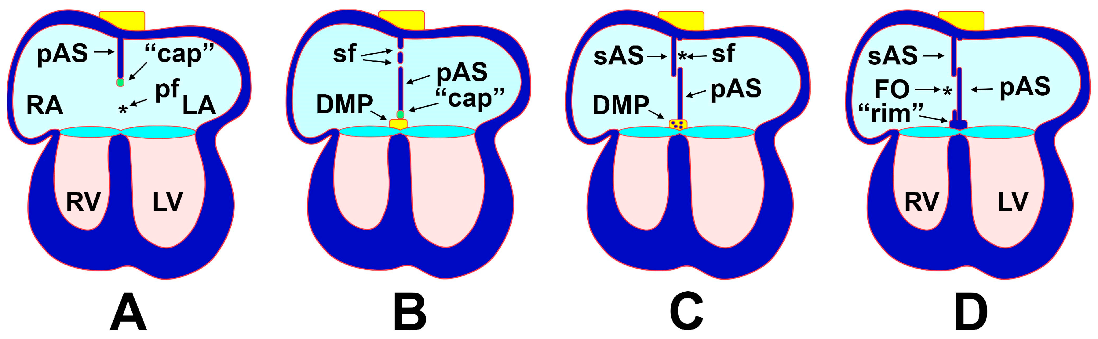
| 23:21, 3 October 2017 | Stages of Atrial Septation.png (file) |  |
5.14 MB | Anatomy of the atrioventricular septation complex in a human four-chambered heart with normal anatomy and in hearts with AVSDs. (A) shows a properly septated heart with a complete atrial and ventricular septum. (B) depicts a heart with an incomplete AV... | 1 |