Uploads by Z3414648
From Embryology
This special page shows all uploaded files.
| Date | Name | Thumbnail | Size | Description | Versions |
|---|---|---|---|---|---|
| 09:18, 24 October 2014 | Hypothyroidism.jpg (file) |  |
565 KB | This image compares a normal thyroid gland (a) anatomy with a hypothyroidism thyroid gland (b). The histology images reveal in the abnormal thyroid gland, there is less thyroid hormone produced due to irregular epithelium on the surface of the follicle... | 1 |
| 13:48, 23 October 2014 | Anterior Pituitary Hormones.jpg (file) |  |
74 KB | This student-drawn diagram is adapted from a diagram found in the research report Gonadotrope and thyrotrope development in the human and mouse anterior pituitary gland published in the Developmental Biology journal. It illustrates the fetal timeline o... | 1 |
| 11:37, 22 October 2014 | Pituitary Development.jpg (file) |  |
41 KB | A cartoon image of the human pituitary gland anatomy showing the Anterior Pituitary (AP) and Posterior Pituitary (NP) separated by the Marginal Zone (MZ). The MZ is surrounded by dilated Rathke's remnant's cysts in orange and labelled RC on the H&E sta... | 1 |
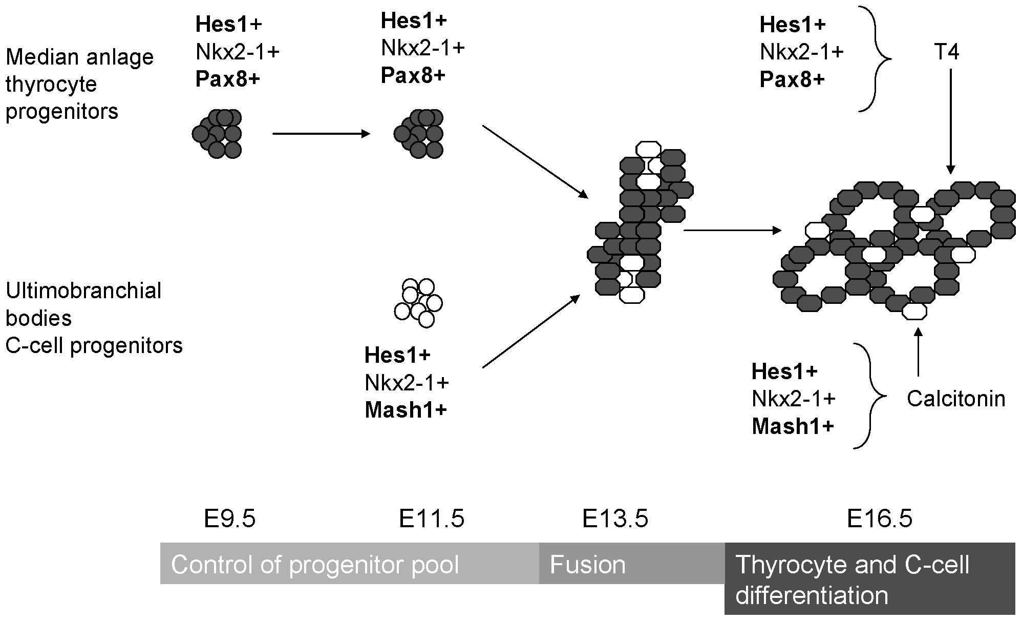
| 13:17, 17 September 2014 | ThyroidDevelopment.png (file) |  |
49 KB | This image summarises the endodermal and mesodermal contribution to the development of the thyroid gland. The progenitor cells are from anterior endoderm and receives buds from left and right endodermal buds covered in mesoderm. <ref name="PMID10.1371/... | 1 |
| 13:35, 19 August 2014 | Cleavage stage embryo.png (file) |  |
7.13 MB | ==Cleavag stage embryo== This image shows the growth receptors involved in the cleavage-stage of embryo development. The red stain is with propidium iodide indicating the nuclei. The green staining areas indicate the ligand-receptor pairs for EGF/EGF... | 1 |