Uploads by Z3333794
From Embryology
This special page shows all uploaded files.
| Date | Name | Thumbnail | Size | Description | Versions |
|---|---|---|---|---|---|
| 10:29, 3 October 2012 | Pharyngeal arch one and two in mice.png (file) |  |
326 KB | The image shows the pharyngeal arches one and two in mice embryo. All three layers ectoderm, mesoderm and endoderm from arch one and two contribute to the formation of the ear. <pubmed>22110697</pubmed> Citation: Diman NYS-G, Remacle S, Bertrand N, Pi | 1 |
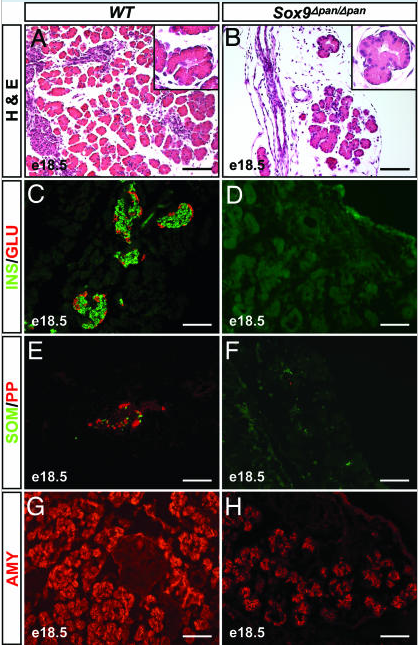
| 21:52, 2 October 2012 | Sox9.png (file) |  |
368 KB | 1 | |
| 15:17, 2 October 2012 | Histology of Inner Ear.png (file) |  |
200 KB | 1 | |
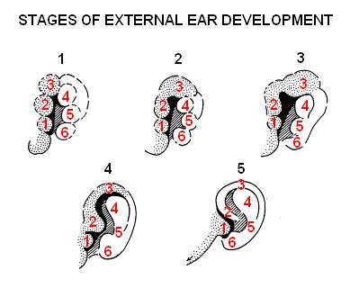
| 11:42, 26 September 2012 | Devt of external ear.JPG (file) |  |
20 KB | 1 | |
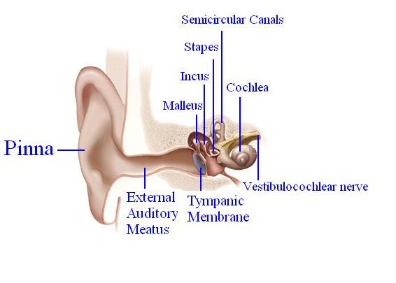
| 12:16, 19 September 2012 | Anatomy of the Ear.JPG (file) |  |
23 KB | '''The image shows the anatomy of adult human ear:''' Outer ear: Pinna, Eaxternal auditory meatus and tympanic membrane Middle ear: Ossicles - Malleus, Incus and Stapes Inner ear: Cochlea containing the Organ of Corti and Semi-circular canals containin | 1 |
| 12:11, 19 September 2012 | Ananomy of Ear.JPG (file) |  |
23 KB | The image shows the anatomy of adult human ear: Outer ear: Pinna, Eaxternal auditory meatus and tympanic membrane Middle ear: Ossicles - Malleus, Incus and Stapes Inner ear: Cochlea containing the Organ of Corti and Semi-circular canals containing semi | 1 |
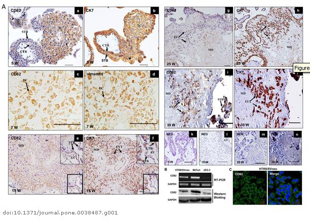
| 13:45, 7 August 2012 | Expression of CD82 in human placental villi and cell lines.JPG (file) |  |
75 KB | Expression of CD82 in human placental villi and cell lines. http://www.plosone.org/article/info%3Adoi%2F10.1371%2Fjournal.pone.0038487 | 1 |
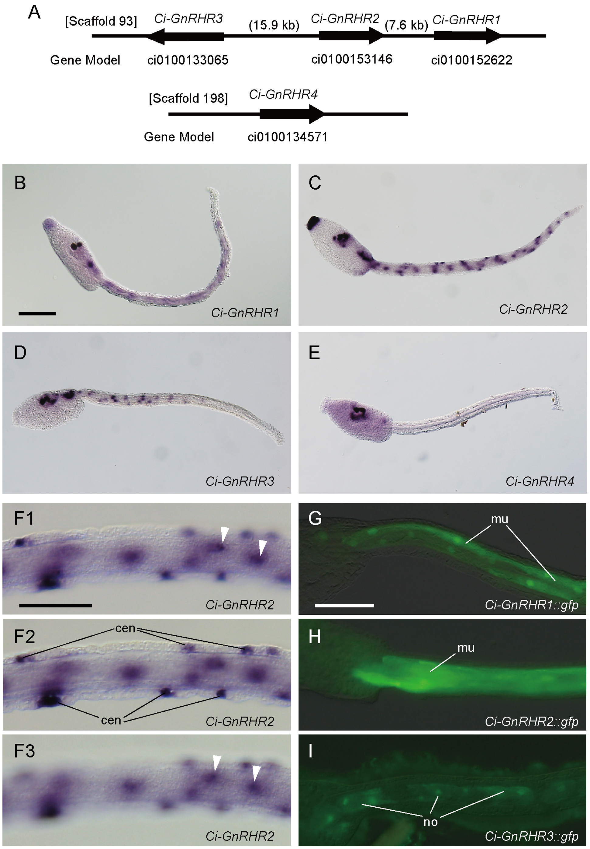
| 11:59, 1 August 2012 | GnRH receptors (GnRHRs) and spatial expression patterns of gnrhr genes.png (file) | _and_spatial_expression_patterns_of_gnrhr_genes.png) |
6.16 MB | http://www.plosone.org/article/info%3Adoi%2F10.1371%2Fjournal.pone.0041955 | 1 |