Uploads by Z3308968
From Embryology
This special page shows all uploaded files.
| Date | Name | Thumbnail | Size | Description | Versions |
|---|---|---|---|---|---|
| 21:36, 9 October 2011 | Migration.jpg (file) |  |
118 KB | The migration of neural crest cells into the mesenchymal tissues of the pharyngeal arches and subsequent differentiation into the bones of the craniofacial skeleton The process of neural crest cell migration and differentiation leads to the development o | 1 |
| 22:47, 2 October 2011 | Identification of Van Der Woude syndrome by lesions on lower lips.jpg (file) |  |
41 KB | 2 | |
| 23:26, 26 September 2011 | Velocardiofacial syndrome with typical facies.jpg (file) |  |
171 KB | ===Reference=== <pubmed>19884681</pubmed>| [http://www.ncbi.nlm.nih.gov/pmc/articles/PMC2825080 PMC2825080] Velocardiofacial syndrome (VCFS) is an autosomal dominant condition which results from a deletion on the long arm of Chromosome 22 in the“q11� | 4 |
| 00:31, 22 September 2011 | Van der Woude syndrome with lower lip pits.jpg (file) |  |
170 KB | Van der Woude syndrome with lower lip pits-Most common syndrome associated with cleft http://www.ncbi.nlm.nih.gov/pmc/articles/PMC2825080/ This is an open-access article distributed under the terms of the Creative Commons Attribution License, which perm | 1 |
| 00:23, 22 September 2011 | Median facial dysplasia.jpg (file) |  |
179 KB | Median facial dysplasia-A facial anomaly associated with cleft http://www.ncbi.nlm.nih.gov/pmc/articles/PMC2825080/ This is an open-access article distributed under the terms of the Creative Commons Attribution License, which permits unrestricted use, d | 1 |
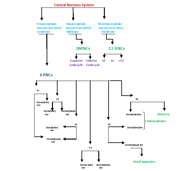
| 22:59, 19 September 2011 | Figure Shows How the CNS is divided to supply different structures.jpg (file) |  |
123 KB | Figure Shows How the CNS is divided to supply different structures The forebrain is made up of six prosomeres. These have designated numbers from caudal to cranial. Each prosemere are subdivided into two tiers, dorsal (alar) and ventral (basal). The alar | 1 |
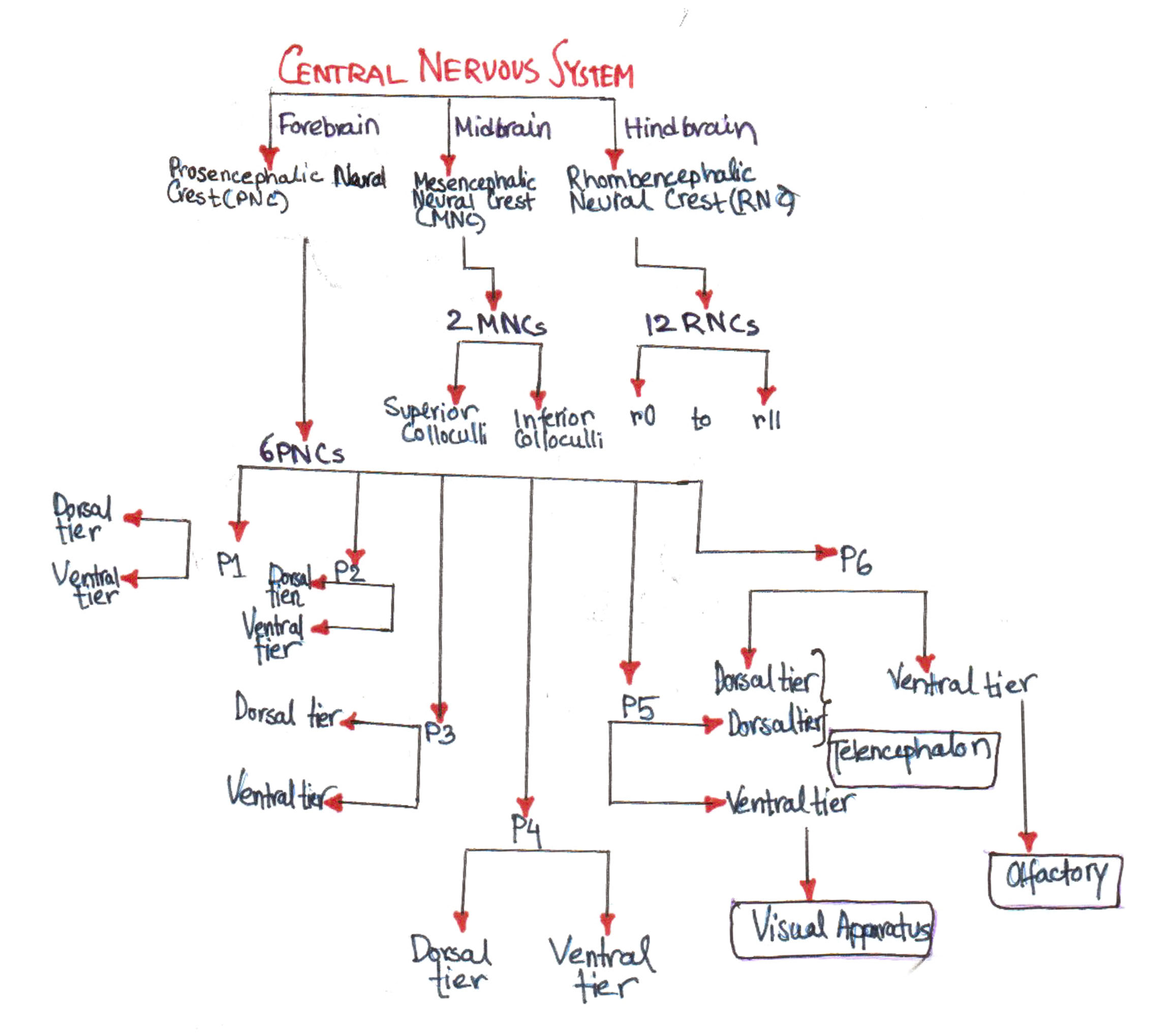
| 12:57, 17 September 2011 | Origin of the craniofacial skeleton-1.jpg (file) |  |
123 KB | The forebrain is made up of six prosomeres. These have designated numbers from caudal to cranial. Each prosemere are subdivided into two tiers, dorsal (alar) and ventral (basal). The alar tiers of p6 and p5 gives rise to the telencephalon while the basal | 1 |
| 23:46, 14 September 2011 | Origin of the craniofacial skeleton.jpg (file) |  |
319 KB | The forebrain is made up of six prosomeres. These have designated numbers from caudal to cranial. Each prosemere are subdivided into two tiers, dorsal (alar) and ventral (basal). The alar tiers of p6 and p5 gives rise to the telencephalon while the basal | 3 |
| 20:26, 12 September 2011 | NeuromericOrganization.jpg (file) |  |
123 KB | The neuromeric organization of the human embryo. Reproduced from Rubenstein, 1994 This is an open-access article distributed under the terms of the Creative Commons Attribution License, which permits unrestricted use, distribution, and reproduction in an | 1 |
| 22:34, 8 September 2011 | Veau-Wardill-Kilner technique of palate repair in a unilateral cleft lip and palate.jpg (file) |  |
165 KB | Figure 2a-d Line diagram showing the Veau-Wardill-Kilner technique of palate repair in a unilateral cleft lip and palate http://www.ncbi.nlm.nih.gov/pmc/articles/PMC2825076/figure/F0002/ This is an open-access article distributed under the terms of the | 1 |
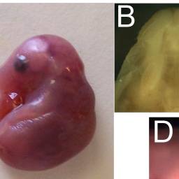
| 12:37, 18 August 2011 | Mice mutants exhibit cleft palate and umbilical hernia.jpg (file) |  |
126 KB | Mice_mutants_exhibit_cleft_palate_and_umbilical_hernia http://www.ncbi.nlm.nih.gov/pmc/articles/PMC2841638/?tool=pubmed Oh et al. This is an open-access article distributed under the terms of the Creative Commons Attribution License, which permits unres | 1 |
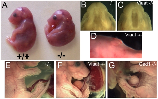
| 12:32, 18 August 2011 | Viaat mutants exhibit cleft palate and umbilical hernia.jpg (file) |  |
7 KB | Viaat mice mutants exhibit cleft palate and umbilical hernia http://www.ncbi.nlm.nih.gov/pmc/articles/PMC2841638/?tool=pubmed Oh et al. This is an open-access article distributed under the terms of the Creative Commons Attribution License, which permits | 1 |
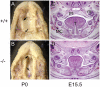
| 10:56, 18 August 2011 | Pone.0014375.g001.gif (file) |  |
9 KB | The recessive ENU-induced csp1 mutation in Prdm16 exhibits cleft secondary palate. http://www.ncbi.nlm.nih.gov/pmc/articles/PMC3002952/?tool=pubmed Bjork et al. This is an open-access article distributed under the terms of the Creative Commons Attributi | 1 |
| 10:53, 18 August 2011 | Photos of fusion or lack of fusion by embryonic palatal shelves in mice.jpg (file) |  |
6 KB | Photos of fusion or lack of fusion by embryonic palatal shelves. The fusion situation of embryonic palatal shelves was observed and photographed. (a) Fused embryonic palatal shelf. (b) Nonfused embryonic palatal shelf. (c) Fused embryonic palatal shelf | 1 |
| 10:42, 18 August 2011 | Histological Comparison of Cleft Palate in Mice.jpg (file) |  |
7 KB | Comparison of cleft palate and omphalocele between VGAT-/- mice and GAD67-/- mice http://www.ncbi.nlm.nih.gov/pmc/articles/PMC3023674/?tool=pubmed This is an Open Access article distributed under the terms of the Creative Commons Attribution License ( | 1 |
| 18:50, 15 August 2011 | Cleft Palate.jpg (file) |  |
71 KB | Copyright Information I, the copyright holder of this work, release this work into the public domain. This applies worldwide. In some countries this may not be legally possible; if so: I grant anyone the right to use this work for any purpose, without any | 1 |