Uploads by Z3252833
From Embryology
This special page shows all uploaded files.
| Date | Name | Thumbnail | Size | Description | Versions |
|---|---|---|---|---|---|
| 20:40, 6 October 2010 | ZHydrophone.jpg (file) |  |
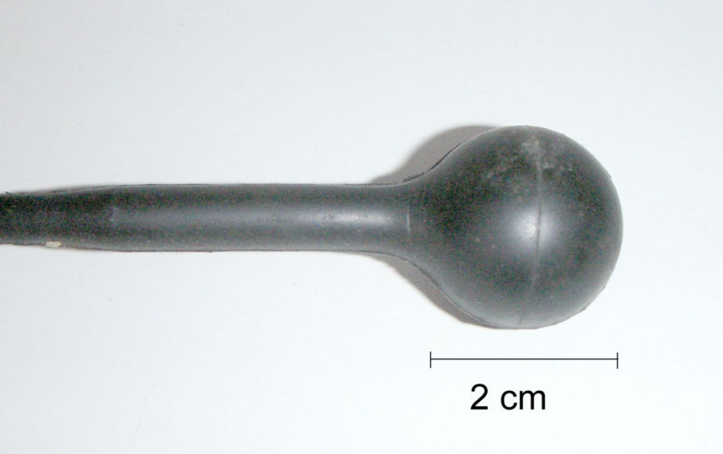
29 KB | ===What am I looking at?=== This is an image of a hydrophone. It is an ultrasonic transducer working on the principle of the piezoelectric effect that is used underwater to send out ultrasound waves in order to try and locate large objects underwater. It | 1 |
| 20:24, 6 October 2010 | ZUltrasound Exam.JPG (file) |  |
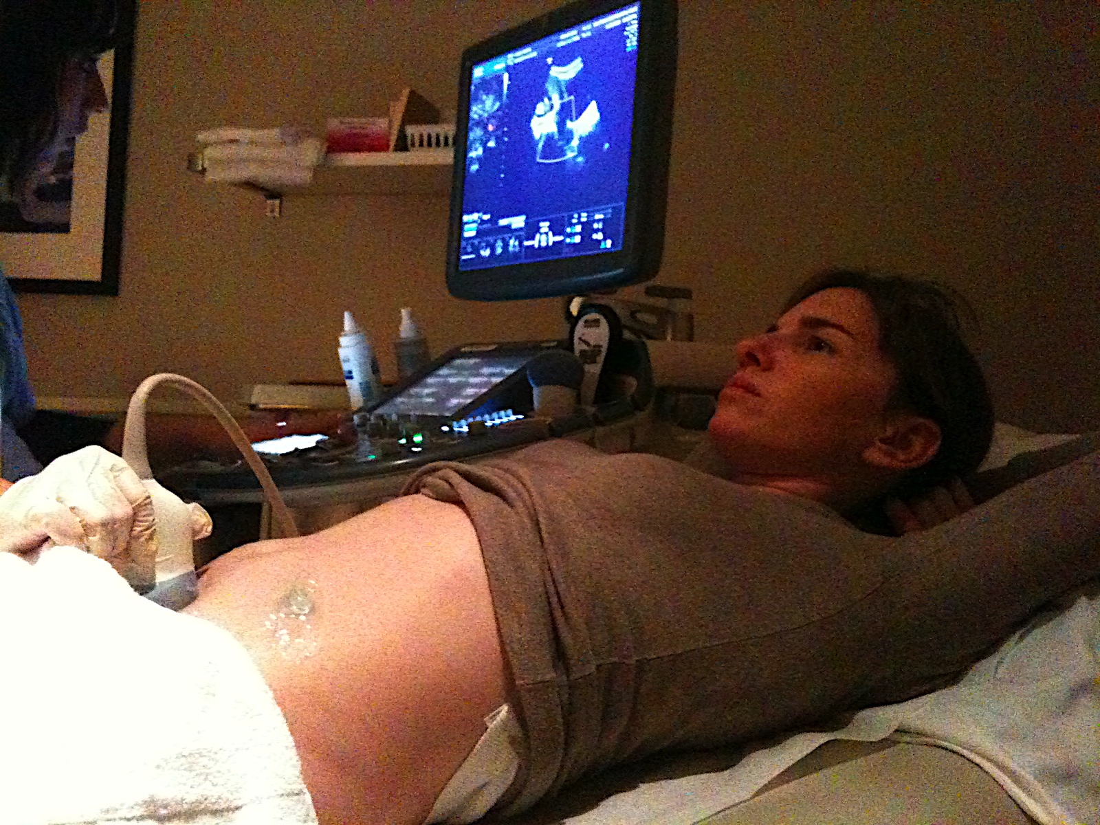
846 KB | ===What am I looking at?=== This is a photograph of a pregnant woman having an ultrasound examination to screen the growing fetus for any overt structural abnormalities. The gel applied to her abdomen allows for a more efficient interface between the tra | 1 |
| 20:10, 6 October 2010 | ZUltrasound System.jpg (file) |  |
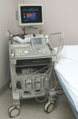
49 KB | ===What am I looking at?=== This is a photograph of a relatively modern ultrasound system, clearly showing the computer component and the monitor that the ultrasound images are displayed upon. ===Image Copyright Information=== Image Author: Daniel W. | 1 |
| 19:38, 26 September 2010 | ZAnencephaly.jpg (file) |  |
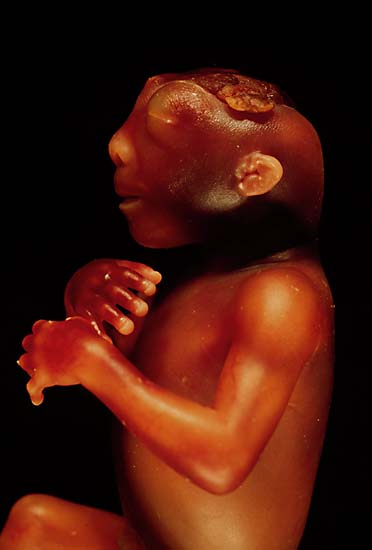
16 KB | ==What am I looking at?== This is a picture of a fetus with anencephaly; a neural tube defect in which the cerebral hemispheres do not develop. ==Image Copyright Information== Image source: http://www.geocities.com/HotSprings/Falls/7780/images /anence | 1 |
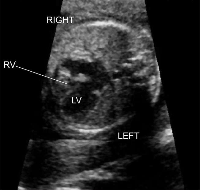
| 17:07, 26 September 2010 | ZPulmonary Atresia.jpg (file) |  |
85 KB | ==What am I looking at?== This is a 2D ultrasound of a fetal heart, showing pulmonary atresia with in intact ventricular septum. This is a four-chamber view of the heart showing that the left ventricle is greater in size than the right ventricle. The rig | 1 |
| 17:24, 12 September 2010 | ZScan Lines.jpg (file) |  |
322 KB | ===Explanation of Diagram=== This diagram is designed to show that a single pulse of ultrasound produces one scan line of information, so many pulses of ultrasound together produce many scan lines and ultimately a more complete cross-sectional image of t | 1 |
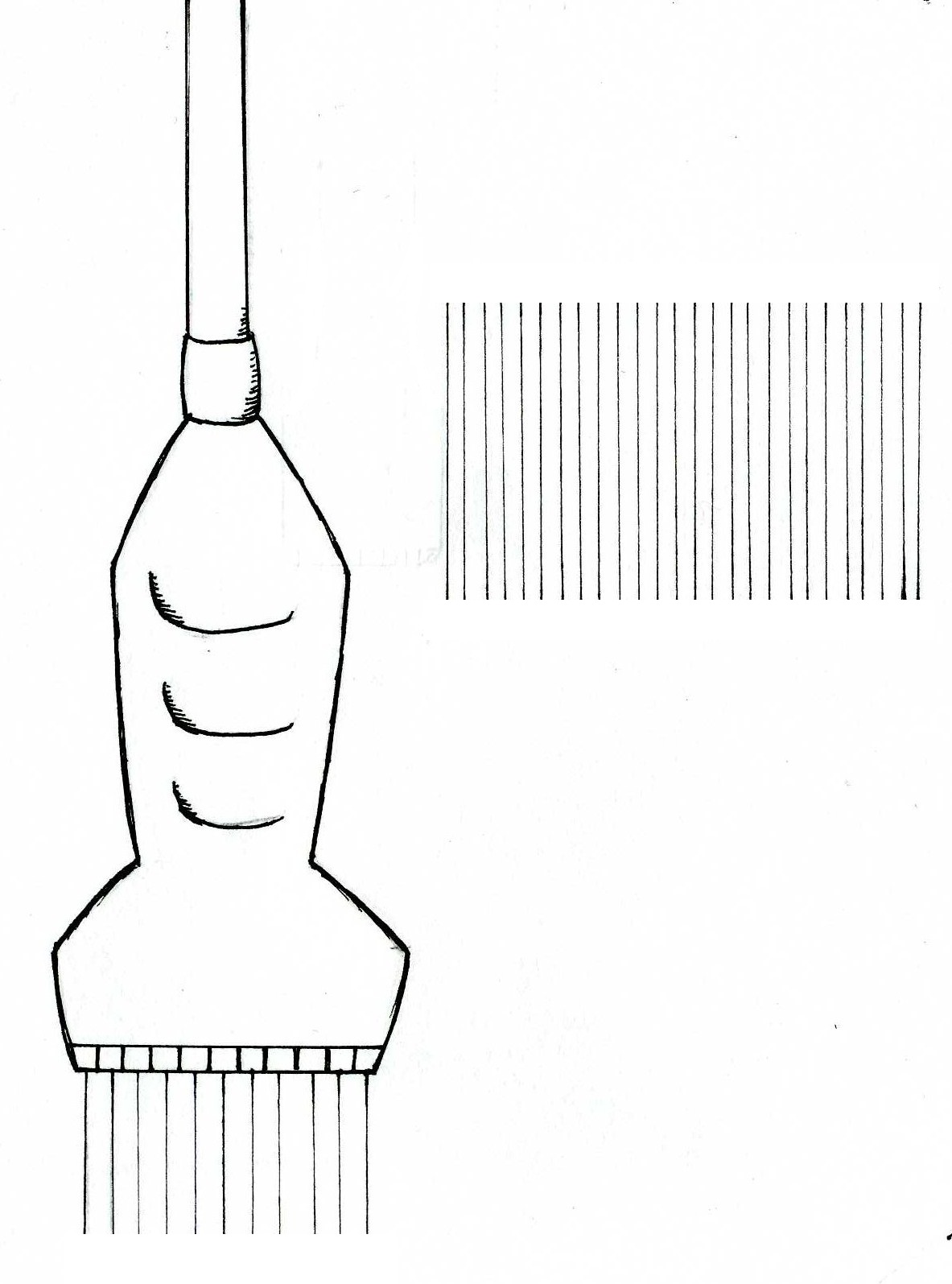
| 17:18, 12 September 2010 | ZPhased Array Transducerki.jpg (file) |  |
214 KB | ===Explanation of Diagram=== This diagram shows a phased array transducer (also called a linear phased array transducer) emitting ultrasound pulses on the left, and the shape of the scan it produces on the right. The illustration of a phased array trans | 1 |
| 17:12, 12 September 2010 | ZConvex Array Transducer.jpg (file) |  |
441 KB | ===Explanation of Diagram=== This diagram shows a curved or convex array transducer emitting ultrasound pulses on the left, and the shape of the scan it produces on the right. The illustration of a convex or curved array transducer on the left demonstr | 1 |
| 16:49, 12 September 2010 | ZLinear Array Transducer.jpg (file) |  |
221 KB | ===Explanation of Diagram=== This diagram shows a linear array transducer emitting ultrasound pulses on the left, and the shape of the scan it produces on the right. The illustration of a linear transducer on the left demonstrates the placement of piez | 1 |
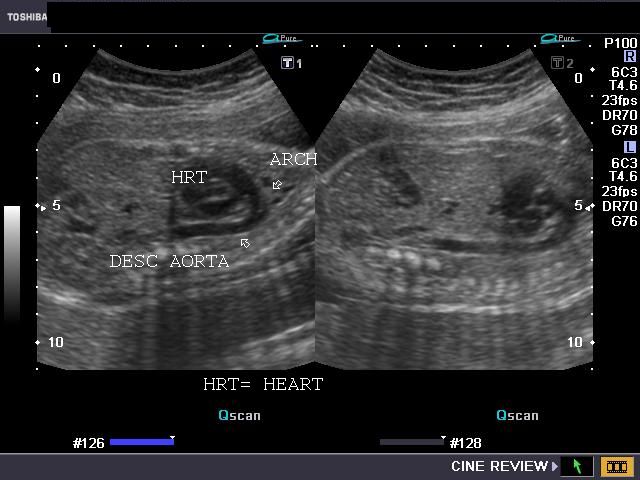
| 19:10, 10 September 2010 | ZUltrasound Image of Fetal Aorta.jpg (file) |  |
47 KB | ===What am I looking at?=== This is a 2D ultrasound image of a fetal chest in sagittal section. Marked with arrows on the image are the fetal heart, aortic arch and descending aorta. ===Image Copyright Information=== Image obtained at: http://www.ul | 1 |
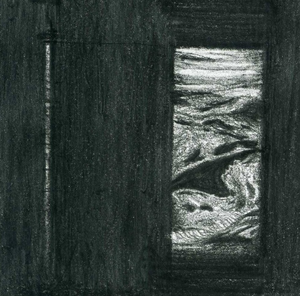
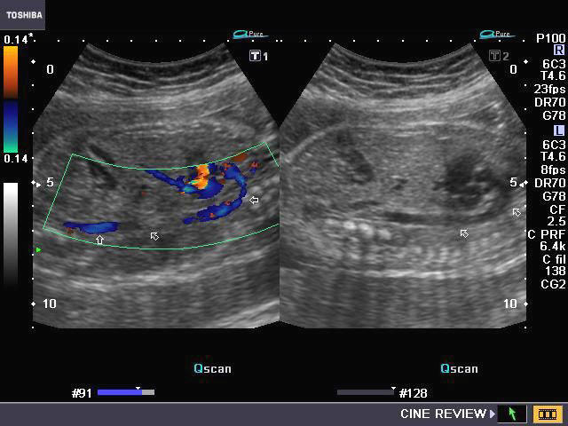
| 18:56, 10 September 2010 | ZDoppler Image of Fetal Aorta.jpg (file) |  |
53 KB | ===What am I looking at?=== This is a colour Doppler ultrasound image depicting a sagittal section of the chest of a fetus. The aortic arch and descending thoracic aorta can be seen extending from the left ventricular outflow tract. Colour is used to | 1 |
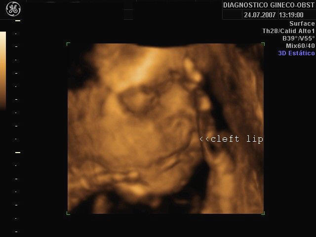
| 17:31, 10 September 2010 | Z3DCleft Lip Picture.jpg (file) |  |
32 KB | ==What am I looking at?== This is a 3D ultrasound image showing a fetus with a cleft lip. The 3D rendering enables the abnormality to be clearly visualised. ==Image Copyright Information== Image obtained at: http://www.ultrasound-images.com/fetal-fa | 1 |