Prize Essay on the Corpus Luteum (1851) Plates
Explanation of the Plates
| Embryology - 19 Apr 2024 |
|---|
| Google Translate - select your language from the list shown below (this will open a new external page) |
|
العربية | català | 中文 | 中國傳統的 | français | Deutsche | עִברִית | हिंदी | bahasa Indonesia | italiano | 日本語 | 한국어 | မြန်မာ | Pilipino | Polskie | português | ਪੰਜਾਬੀ ਦੇ | Română | русский | Español | Swahili | Svensk | ไทย | Türkçe | اردو | ייִדיש | Tiếng Việt These external translations are automated and may not be accurate. (More? About Translations) |
Dalton JC. Prize essay on the corpus luteum of menstruation and pregnancy. (1851) Philadelphia: T.K. and P.G. Collins.
| Online Editor |
|---|
| This historic 1851 paper by Dalton is a very early historic description of the corpus luteum.
|
| Historic Disclaimer - information about historic embryology pages |
|---|
| Pages where the terms "Historic" (textbooks, papers, people, recommendations) appear on this site, and sections within pages where this disclaimer appears, indicate that the content and scientific understanding are specific to the time of publication. This means that while some scientific descriptions are still accurate, the terminology and interpretation of the developmental mechanisms reflect the understanding at the time of original publication and those of the preceding periods, these terms, interpretations and recommendations may not reflect our current scientific understanding. (More? Embryology History | Historic Embryology Papers) |
| Corpus Luteum 1851: Part 1 - Corpus Luteum of Menstruation | Part 2 - Corpus Luteum of Pregnancy | Part 3 - Observations on Animals | Plates |
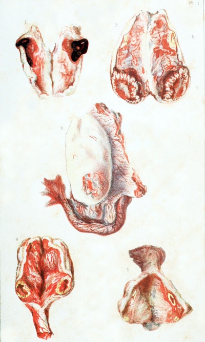
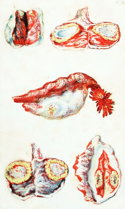
N. B. — All the drawings were taken from nature, and are of the natural size (in the original article). The figures in the first two plates are all taken from the human subject.
Plate I
| Fig. 1. Graafian vesicle, recently ruptured and filled with blood. (Obs. 1.)
Fig. 3. The same ovary cut open; showing the corpus luteum of menstruation three weeks old.
Fig. 4. Corpus luteum of menstruation four weeks old. (Obs. 9.)
|

|
Plate II
| Fig. 1. Corpus luteum of menstruation nine weeks old. (Obs. 11.)
|

|
Plate III
Fig. 1. Ovary of an unimpregnated cow. (Obs. 20.)
a. Old corpus luteum seen externally.
b. New corpus luteum seen externally.
Fig. 2. The same ovary cut open.
a. Old corpus luteum.
b. Ruptured Graafian vesicle in process of transformation into a corpus luteum.
Fig. 3. Corpus luteiim of the unimpregnated cow at its maximum of development. External view. (Obs. 21.)
Fig. 4. Same as the above. Internal view.
Fig. 5. Corpus luteum of the unimpregnated cow beginning to retrograde. External view. (Obs. 22.)
Plate IV
Fig. 1. Same as the above. Internal view.
Fig. 2. Corpus luteum of a cow about three and a half months pregnant. (Obs. 23.)
Fig. 3. Corpus luteum of a cow eight months pregnant. (Obs. 26.)
Fig. 4. Corpus luteum of the unimpregnated ewe at its maximum of development. a. External view. b. Internal view.
Fig. 5. a. Ovary of an unimpregnated ewe ; showing the aperture of a recentlyruptured Graafian vesicle, and the prominence of the last corpus luteum. b. The same ovary cut open ; showing the last corpus luteum.
Fig. 6. a. Ovary of a ewe, the uterus of which contained a foetus eight and a half inches in length. b. The same ovary cut open.
| Embryology - 19 Apr 2024 |
|---|
| Google Translate - select your language from the list shown below (this will open a new external page) |
|
العربية | català | 中文 | 中國傳統的 | français | Deutsche | עִברִית | हिंदी | bahasa Indonesia | italiano | 日本語 | 한국어 | မြန်မာ | Pilipino | Polskie | português | ਪੰਜਾਬੀ ਦੇ | Română | русский | Español | Swahili | Svensk | ไทย | Türkçe | اردو | ייִדיש | Tiếng Việt These external translations are automated and may not be accurate. (More? About Translations) |
Dalton JC. Prize essay on the corpus luteum of menstruation and pregnancy. (1851) Philadelphia: T.K. and P.G. Collins.
| Online Editor | ||||
|---|---|---|---|---|
| This historic 1851 paper by Dalton is a very early historic description of the corpus luteum.
|
| Historic Disclaimer - information about historic embryology pages |
|---|
| Pages where the terms "Historic" (textbooks, papers, people, recommendations) appear on this site, and sections within pages where this disclaimer appears, indicate that the content and scientific understanding are specific to the time of publication. This means that while some scientific descriptions are still accurate, the terminology and interpretation of the developmental mechanisms reflect the understanding at the time of original publication and those of the preceding periods, these terms, interpretations and recommendations may not reflect our current scientific understanding. (More? Embryology History | Historic Embryology Papers) |
| Corpus Luteum 1851: Part 1 - Corpus Luteum of Menstruation | Part 2 - Corpus Luteum of Pregnancy | Part 3 - Observations on Animals | Plates |

