Paper - On the development of the early human ovum, with special reference to the trophoblast of the previllous stage (1944)
| Embryology - 24 Apr 2024 |
|---|
| Google Translate - select your language from the list shown below (this will open a new external page) |
|
العربية | català | 中文 | 中國傳統的 | français | Deutsche | עִברִית | हिंदी | bahasa Indonesia | italiano | 日本語 | 한국어 | မြန်မာ | Pilipino | Polskie | português | ਪੰਜਾਬੀ ਦੇ | Română | русский | Español | Swahili | Svensk | ไทย | Türkçe | اردو | ייִדיש | Tiếng Việt These external translations are automated and may not be accurate. (More? About Translations) |
Hertig AT. and Rock J. On the development of the early human ovum, with special reference to the trophoblast of the previllous stage: A description of a normal and 5 pathologic human ova. (1944) Amer. J. Obstet Gynecol., 47: 149-184.
| Online Editor |
|---|

See also:
Hertig AT. and Rock J. On a normal human ovum not over 7.5 days of age. (1945) Anat. Rec. 91: 281. Hertig AT. and Rock J. On a normal ovum of approximately 9 to 10 days of age. (1945) Anat. Rec. 91: 281. Hertig AT. and Rock J. On a human blastula recovered from the uterine cavity 4 days after ovulation. (1946) J Gerontol. 1(1): 96-117.
|
| Historic Disclaimer - information about historic embryology pages |
|---|
|
On the Development of the Early Human Ovum, with Special Reference to the Trophoblast of the Previllous Stage: A Description of a Normal and 5 Pathologic Human Ova
|

|
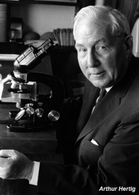
| Arthur Tremain Hertig (1904-1990) | John Charles Rock (1890-1984) |
Abstract
A series of 5 previllous and 2 villous normal human ova, ranging from 7.5 to 16.5 days in developmental age, shows that the human blastocyst implants on the posterior wall, probably during the late sixth or nearly seventh day of its development, on endometrium that may range from the eighteenth to the twenty-third day of its development. Actually there are no precise data on the time of implantation, since the youngest specimen, and therefore the most critical one with respect to this process, is already implanted. The figures given (late sixth or early seventh day) are deduced on the basis of this youngest specimen. Even younger ova must be secured in order to determine the actual time of implantation.
Trophoblast proliferates at the site of implantation which, at first, consists of solid cytotrophoblast and syncytiotrophoblast. The latter becomes vacuolated on the eighth day to develop lacunae for the reception of maternal blood on about the eleventh day. The chorionic villi begin to form as cytotrophoblastic masses on the twelfth to thirteenth day and grow peripherally along the syncytiotrophoblastic framework, ultimately coalescing peripherally to displace the syncytiotrophoblast, except the portion lining the intervillous space. Remnants of the desquamated syncytiotrophoblast are encountered in the placental site as giant cells.
A series of 5 abnormal previllous ova, the developmental ages of which range from approximately the eleventh to the fourteenth day, but which are difficult to interpret accurately because of their abnormality, shows a variety of conditions ranging from shallow implantation of an otherwise normal ovum, through extreme hypoplasia of the trophoblast, to complete absence of the embryonic mass. The pathologic ova were all found on the anterior wall of the uterus.
Cite this page: Hill, M.A. (2024, April 24) Embryology Paper - On the development of the early human ovum, with special reference to the trophoblast of the previllous stage (1944). Retrieved from https://embryology.med.unsw.edu.au/embryology/index.php/Paper_-_On_the_development_of_the_early_human_ovum,_with_special_reference_to_the_trophoblast_of_the_previllous_stage_(1944)
- © Dr Mark Hill 2024, UNSW Embryology ISBN: 978 0 7334 2609 4 - UNSW CRICOS Provider Code No. 00098G