File:Zebrafish enveloping layer SEM01.jpg
Original file (1,200 × 447 pixels, file size: 150 KB, MIME type: image/jpeg)
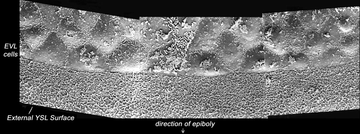
Zebrafish enveloping layer (day 1) SEM
Scanning EM of a 24 hr (prim-5) zebrafish embryo.
- Links: Image - day 1 | Image - brain fold | Image - myotomes | Image - trunk | Image - trunk | Image - perichordal sheath | Image - enveloping layer | Image - enveloping layer | Zebrafish Development | Scanning Electron Microscopy
Image Source: Scanning electron micrographs of the Zebrafish embryos are reproduced with the permission of Associate Professor Bryan Crawford, Department of Biology, University of New Brunswick.
Reference
<pubmed>15602926</pubmed>
Specimens were chemically fixed critically point dried, and sputter coated with gold/palladium. This image is part of a series taken by Bryan Crawford while he was at the University of Washington. They are part of the Zebrafish--The Living Laboratory CD made available by Mark Cooper and described in Methods in Cell Biology Volume 77, 2004, Pages 439-457.
Copyright
Licensing: Attribution Non-Commercial Share Alike:This image is licensed under a Creative Commons Attribution, Non-Commercial Share Alike License.
File history
Click on a date/time to view the file as it appeared at that time.
| Date/Time | Thumbnail | Dimensions | User | Comment | |
|---|---|---|---|---|---|
| current | 14:29, 12 December 2014 | 1,200 × 447 (150 KB) | Z8600021 (talk | contribs) |
You cannot overwrite this file.
File usage
The following page uses this file: