File:Zebrafish- bone growth 01.jpg
Original file (634 × 1,000 pixels, file size: 110 KB, MIME type: image/jpeg)
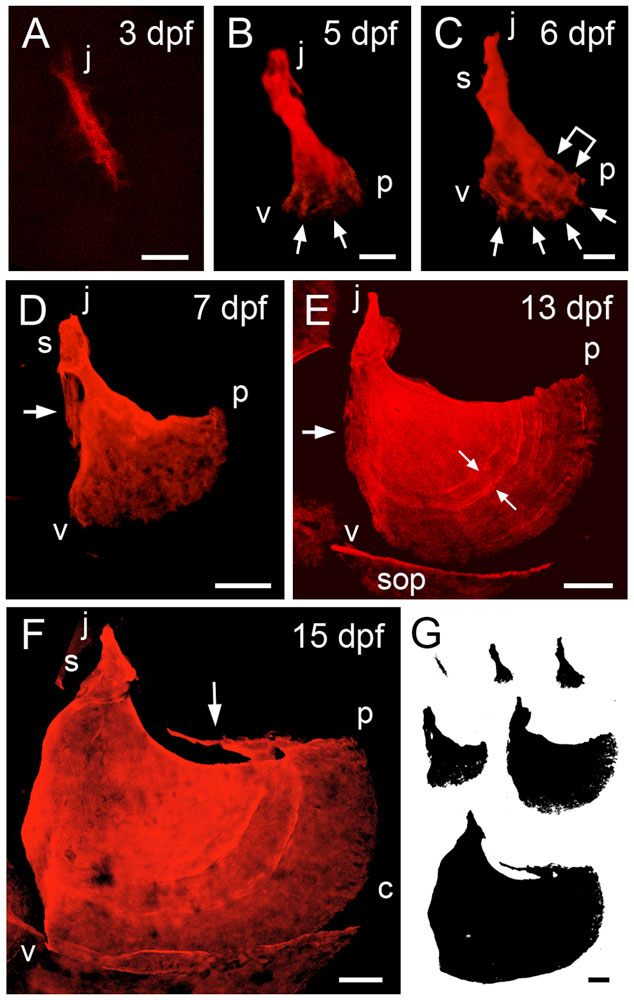
Time-course of shape changes and growth of the opercle in live, developing zebrafish larvae
A-F: Confocal projections made from z-stacks of images of live preparations vitally stained with Alizarin Red S. G: Silhouettes of the same bones scaled to the same final magnification to illustrate the amount of overall bone growth. Left-side views with dorsal approximately to the top and anterior to the left. The same orientations are used in all of the figures for this paper.
(A) The Op initially ossifies as a linear bony spur. The more dorsal or j (‘joint’) end is adjacent to the hyosymplectic cartilage (not shown). In occasional preparations we first see the Op as just a spot of bone at what will become the j end.
(B, C) Early fan-shaped Ops at 5 and 6 dpf, with three apices j (joint apex), v (ventral) and p (posterior). We use vj, vp, and jp to describe the bone edges between these apices. The j end of the element has elaborated a joint socket (s) component of the ball-and-socket articulation the Op makes with the hyosymplectic cartilage (for anatomy see [16]). The posterior-ventral end has broadened to form a new vp edge, by developing small, secondary spurs (arrows), with intervening thinly-mineralized veils. The linked arrow in C shows the first indication of incremental bands.
(D) The fan shape is expanded at 7 dpf by differential elongation of the vp edge, relative to the other two. This vp elongation continues throughout the larval period. A new veil is evident along the vj (anterior) edge (arrow).
(E) The vj veil is still evident at 13 dpf (arrow). A new dorsally pointing spur has appeared at the j apex, the site of attachment of the dilator operculi muscle. Incremental bands are visible in the matrix (e.g. at the small arrow pair). The image includes portions of neighboring ossifications present at this stage, including the subopercle (sop).
(F) By 15 dpf curvature of the vp edge has locally increased at one region (c). The subopercle overlaps the Op vp edge ventrally. The dorsal jp edge, where the levator operculi muscle attaches, has developed a new veil.
Scale bars: 20 µm in A-C, 50 µm in D-G.
Original File name: Figure 1. Journal-1.pone.0009475.g001.jpg
doi:10.1371/journal.pone.0009475.g001
Reference
Copyright: © 2010 Kimmel et al. This is an open-access article distributed under the terms of the Creative Commons Attribution License, which permits unrestricted use, distribution, and reproduction in any medium, provided the original author and source are credited.
File history
Click on a date/time to view the file as it appeared at that time.
| Date/Time | Thumbnail | Dimensions | User | Comment | |
|---|---|---|---|---|---|
| current | 06:58, 22 November 2010 | | 634 × 1,000 (110 KB) | S8600021 (talk | contribs) | ==Time-course of shape changes and growth of the opercle in live, developing zebrafish larvae== A-F: Confocal projections made from z-stacks of images of live preparations vitally stained with Alizarin Red S. G: Silhouettes of the same bones scaled to th |
You cannot overwrite this file.
File usage
The following page uses this file: