File:Zamboni1966 fig06.jpg
Original file (1,280 × 1,158 pixels, file size: 419 KB, MIME type: image/jpeg)
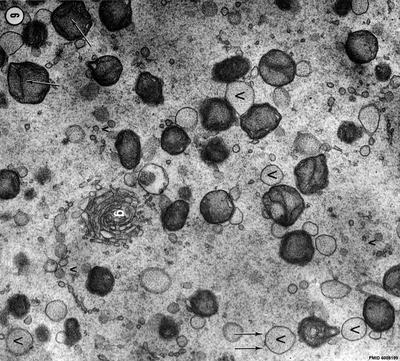
Fig. 6 Region of the ooplasm of the penetrated ovum with typical complement of organelles
The mitochondria are numerous and mostly spheroidal. Two mitochondria with archlike arrangement of the inner cristae are indicated by the single arrows. The ER is present in the form of large (V) and small vesicles (:2). The large vesicles are usually associated with mitochondria. The small vesicles are thought to originate by budding from the large ER vesicles (see figs. 6 to 8). The double arrow indicates a large vesicle with a few ribosomes attached to its limiting membrane. g, Golgi complex. X 29,000.
| Historic Disclaimer - information about historic embryology pages |
|---|
 |
Links: Zamboni 1966 | Historic Embryology Papers
Reference
Zamboni L. Mishell DR. Jr. Bell JH. snd Baca M. Fine structure of the human ovum in the pronuclear stage. (1966) J. Cell Biol, 30: 579-600. PMID 6008199
Copyright
Rockefeller University Press - Copyright Policy This article is distributed under the terms of an Attribution–Noncommercial–Share Alike–No Mirror Sites license for the first six months after the publication date (see http://www.jcb.org/misc/terms.shtml). After six months it is available under a Creative Commons License (Attribution–Noncommercial–Share Alike 4.0 Unported license, as described at https://creativecommons.org/licenses/by-nc-sa/4.0/ ). (More? Help:Copyright Tutorial)
Cite this page: Hill, M.A. (2024, April 23) Embryology Zamboni1966 fig06.jpg. Retrieved from https://embryology.med.unsw.edu.au/embryology/index.php/File:Zamboni1966_fig06.jpg
- © Dr Mark Hill 2024, UNSW Embryology ISBN: 978 0 7334 2609 4 - UNSW CRICOS Provider Code No. 00098G
File history
Click on a date/time to view the file as it appeared at that time.
| Date/Time | Thumbnail | Dimensions | User | Comment | |
|---|---|---|---|---|---|
| current | 14:26, 23 April 2016 | | 1,280 × 1,158 (419 KB) | Z8600021 (talk | contribs) |
You cannot overwrite this file.
File usage
The following page uses this file: