File:ZLinear Array Transducer.jpg
Original file (1,183 × 1,595 pixels, file size: 221 KB, MIME type: image/jpeg)
Explanation of Diagram
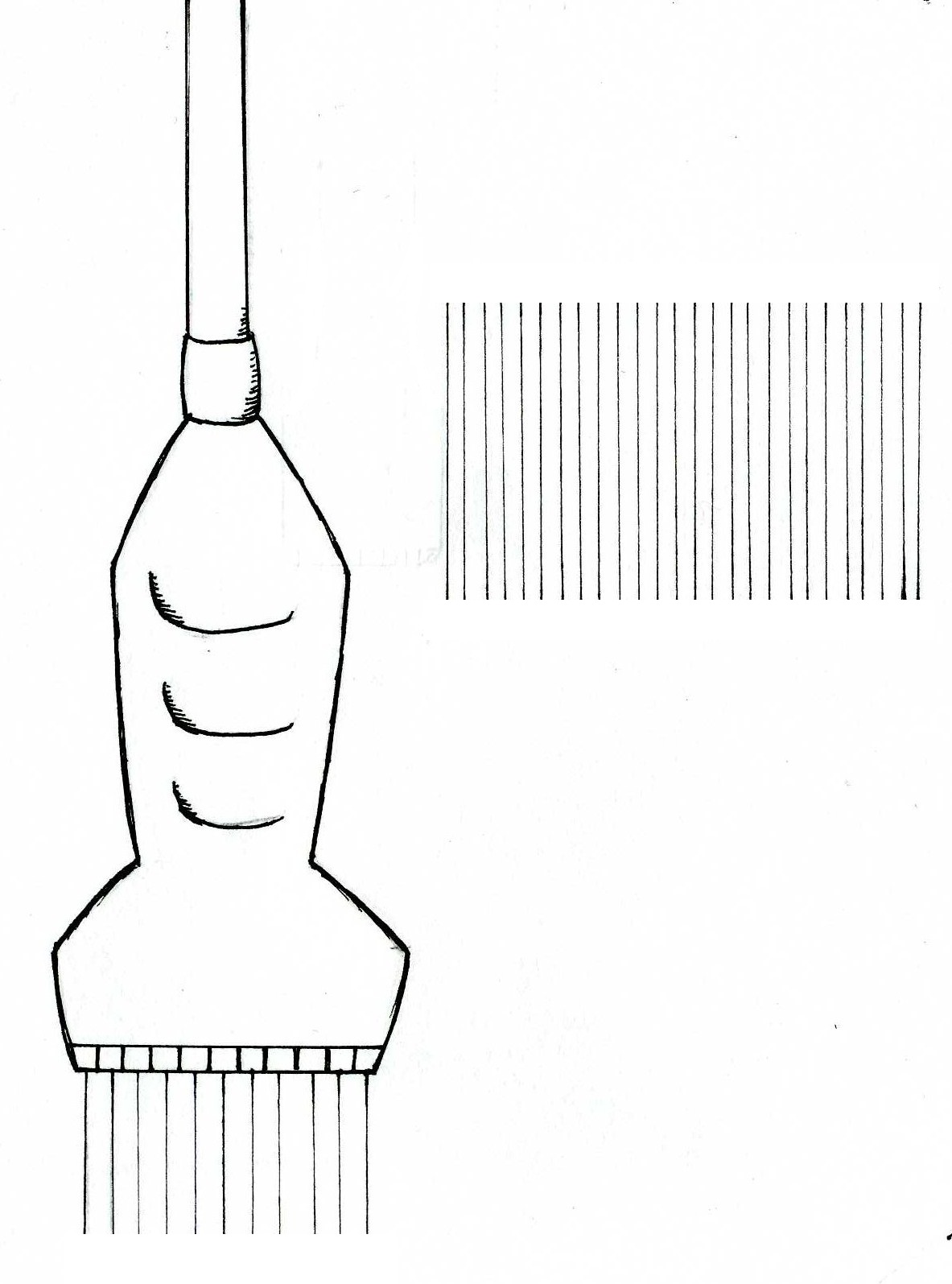
This student-drawn diagram shows a linear array transducer emitting ultrasound pulses on the left, and the shape of the scan it produces on the right.
The illustration of a linear transducer on the left demonstrates the placement of piezoelectric elements in a linear array transducer – side by side, with pulses being emitted vertically and in parallel lines. A complete name for this transducer is the linear sequenced array transducer (‘sequenced’ meaning that it automatically emits pulses in sequence to form a real-time image of the patient).
The illustration on the right indicates the shape of the image produced, termed a linear or rectangular scan.
- Note - This image was originally uploaded as part of an undergraduate science student project and may contain inaccuracies in either description or acknowledgements. Students have been advised in writing concerning the reuse of content and may accidentally have misunderstood the original terms of use. If image reuse on this non-commercial educational site infringes your existing copyright, please contact the site editor for immediate removal.
Image Copyright Information
Illustration by z3252833.
Beginning six months after publication, I, z3252833, grant the public the non-exclusive right to copy, distribute, or display the Work under a Creative Commons Attribution-Noncommercial-Share Alike 3.0 Unported license, as described at http://creativecommons.org/licenses/by-nc-sa/3.0/ and http://creativecommons.org/licenses/by-nc-sa/3.0/legalcode.
References
Kremkali, F.W. (2006) Diagnostic Ultrasound Principles and Instruments (7th ed.) St Louis: Saunders Elsevier. pp 66-67
File history
Click on a date/time to view the file as it appeared at that time.
| Date/Time | Thumbnail | Dimensions | User | Comment | |
|---|---|---|---|---|---|
| current | 16:49, 12 September 2010 | | 1,183 × 1,595 (221 KB) | Z3252833 (talk | contribs) | ===Explanation of Diagram=== This diagram shows a linear array transducer emitting ultrasound pulses on the left, and the shape of the scan it produces on the right. The illustration of a linear transducer on the left demonstrates the placement of piez |
You cannot overwrite this file.
File usage
The following page uses this file: