File:XMRV-infected prostate cells.png
XMRV-infected_prostate_cells.png (485 × 599 pixels, file size: 528 KB, MIME type: image/png)
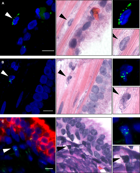
Figure 8. Characterization of XMRV-Infected Prostatic Cells by FISH and FISH/Immunofluorescence
Using a tissue microarray, prostatic tumor tissue sections from QQ case VP62 were analyzed by FISH (green) using DNA probes derived from XMRV VP35 (left panels). Nuclei were counterstained with DAPI. The same sections were then visualized by H&E staining (middle panels). Arrows indicate FISH-positive cells, and their enlarged FISH and H&E images are shown in the top right and bottom right panels, respectively. Scale bars are 10 μm.
(A) A stromal fibroblast.
(B) A dividing stromal cell.
(C) A stromal hematopoietic cell. The section was concomitantly stained for XMRV by FISH (green) and cytokeratin AE1/AE3 by immunofluorescence (red).
Identification of a novel Gammaretrovirus in prostate tumors of patients homozygous for R462Q RNASEL variant. Urisman A, Molinaro RJ, Fischer N, Plummer SJ, Casey G, Klein EA, Malathi K, Magi-Galluzzi C, Tubbs RR, Ganem D, Silverman RH, DeRisi JL. PLoS Pathog. 2006 Mar;2(3):e25. Epub 2006 Mar 31. PMID: 16609730 | PLoS Pathog
Citation: Urisman A, Molinaro RJ, Fischer N, Plummer SJ, Casey G, et al. (2006) Identification of a Novel Gammaretrovirus in Prostate Tumors of Patients Homozygous for R462Q RNASEL Variant. PLoS Pathog 2(3): e25. doi:10.1371/journal.ppat.0020025
Editor: Susan Ross, University of Pennsylvania School of Medicine, United States of America
Received: November 29, 2005; Accepted: February 23, 2006; Published: March 31, 2006
Copyright: © 2006 Urisman et al. This is an open-access article distributed under the terms of the Creative Commons Attribution License, which permits unrestricted use, distribution, and reproduction in any medium, provided the original author and source are credited.
File history
Click on a date/time to view the file as it appeared at that time.
| Date/Time | Thumbnail | Dimensions | User | Comment | |
|---|---|---|---|---|---|
| current | 10:13, 9 October 2009 |  | 485 × 599 (528 KB) | S8600021 (talk | contribs) | Figure 8. Characterization of XMRV-Infected Prostatic Cells by FISH and FISH/Immunofluorescence Using a tissue microarray, prostatic tumor tissue sections from QQ case VP62 were analyzed by FISH (green) using DNA probes derived from XMRV VP35 (left panel |
You cannot overwrite this file.
File usage
There are no pages that use this file.
