File:Wyburn1937 text-fig03.jpg
Original file (1,629 × 700 pixels, file size: 160 KB, MIME type: image/jpeg)
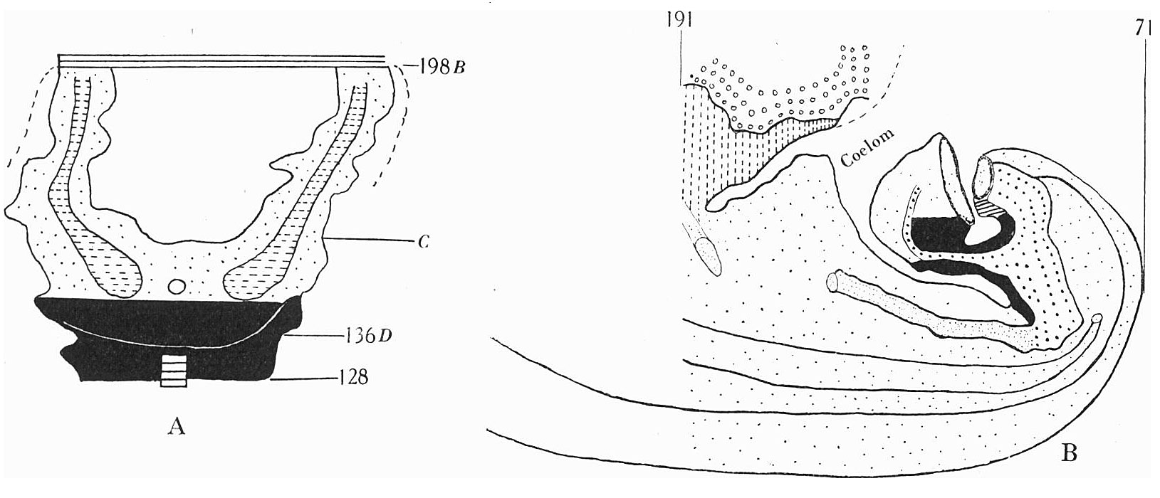
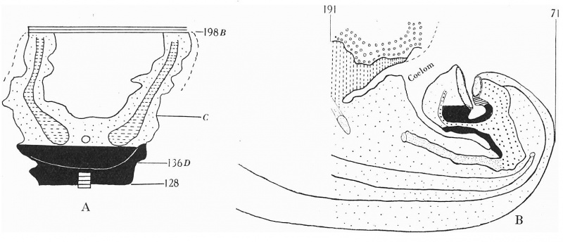
Text-fig. 3. Embryo 4.5 mm (McIntyre II)
X50. The numbers refer to the sections.
A. Reconstruction of the ventral surface from septum transversum to cloacal membrane. The tail is represented straightened out.
B. Reconstruction of median sagittal section (see Pl. VI, fig. 21). The yolk sac is not shown.
Description in text. Gut — fine stippling; cloaca and allantois — large dots; dense mesoderm — black; cloacal membrane — horizontal lines; allantoic vessels ~ interrupted horizontal lines; septum transversum — interrupted vertical lines; liver — small circles. The interval between B and 0 is the communication between extra- and intra-embryonic coelom. D = the level of the caudal attachment of the body stalk.
| Historic Disclaimer - information about historic embryology pages |
|---|
 |
- Links: Text-fig.1. Embryo McIntyre I | Text-fig.2. Embryo 2.4 mm | Text-fig.3. Embryo 4.5 mm McIntyre II | Text-fig.4. Embryo 7 mm | Text-fig.5. Embryo 12.5 mm | Text-fig.6. Embryo 13 mm | Text-fig.7. Embryo 14 mm | Text-fig.8. Embryo 16.1 mm | Text-fig.9. Embryo 23 mm | Text-fig.10. Embryo 30 mm | Text-fig.1. Embryo McIntyre I | Plate 1 | Plate 2 | Plate 3 | Plate 4 | Plate 5 | Plate 6 | Plate 7 | Plate 8
Reference
Wyburn GM. The development of the infra-umbilical portion of the abdominal wall, with remarks on the aetiology of ectopia vesicae. (1937) J Anat. 71(2): 201-31. PMID 17104636
Cite this page: Hill, M.A. (2024, April 18) Embryology Wyburn1937 text-fig03.jpg. Retrieved from https://embryology.med.unsw.edu.au/embryology/index.php/File:Wyburn1937_text-fig03.jpg
- © Dr Mark Hill 2024, UNSW Embryology ISBN: 978 0 7334 2609 4 - UNSW CRICOS Provider Code No. 00098G
File history
Click on a date/time to view the file as it appeared at that time.
| Date/Time | Thumbnail | Dimensions | User | Comment | |
|---|---|---|---|---|---|
| current | 17:28, 15 August 2015 | | 1,629 × 700 (160 KB) | Z8600021 (talk | contribs) | |
| 17:24, 15 August 2015 | 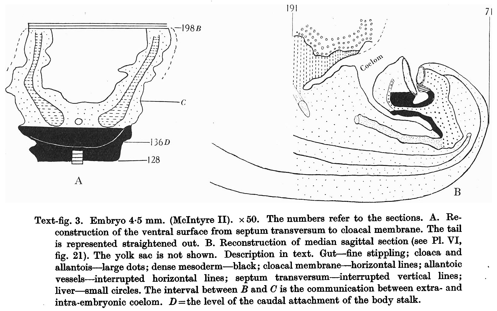 | 1,645 × 1,031 (315 KB) | Z8600021 (talk | contribs) |
You cannot overwrite this file.
File usage
The following page uses this file: