File:Wislocki1920 plate 4.jpg

Original file (935 × 1,000 pixels, file size: 199 KB, MIME type: image/jpeg)
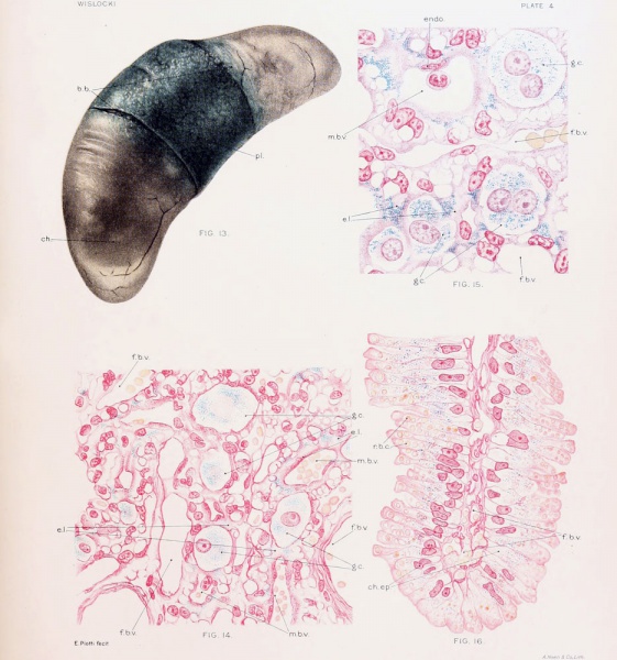
Plate 4. Cat Fetus and Placenta
Fig. 13. Cat fetus measuring 6.8 cm surrounded by enraptured membranesShowing the coloration of the placenta and chorion after repeated injection of trypan-blue into the maternal circulation. Note that the chorion over the poles of the fetus is unstained. The allantoic and amniotic fluids do not contain a trace of dye and the fetus is unstained. Fig. 14. Placenta of cat nearly full termAfter repeated injection of trypan-blue into the maternal blood stream, showing the distribution of dye in the chorionic epithelium. Fig. 15. Placenta of a vitally stained cat nearly full termShowing several multimiclear giant cells of the chorionic ectoderm filled with particles of trypan-blue. Fig. 16. Section of the "brown border" of the placenta of a catNearly full term, showing the absorption of erythrocytes and of trypan-blue by the chorionic epithelium. |
|
| Historic Disclaimer - information about historic embryology pages |
|---|
| Pages where the terms "Historic" (textbooks, papers, people, recommendations) appear on this site, and sections within pages where this disclaimer appears, indicate that the content and scientific understanding are specific to the time of publication. This means that while some scientific descriptions are still accurate, the terminology and interpretation of the developmental mechanisms reflect the understanding at the time of original publication and those of the preceding periods, these terms, interpretations and recommendations may not reflect our current scientific understanding. (More? Embryology History | Historic Embryology Papers) |
File history
Click on a date/time to view the file as it appeared at that time.
| Date/Time | Thumbnail | Dimensions | User | Comment | |
|---|---|---|---|---|---|
| current | 17:45, 27 December 2012 |  | 935 × 1,000 (199 KB) | Z8600021 (talk | contribs) |
You cannot overwrite this file.
File usage
The following 2 pages use this file:
