File:Wislocki1920 plate 1.jpg

Original file (1,145 × 1,200 pixels, file size: 173 KB, MIME type: image/jpeg)
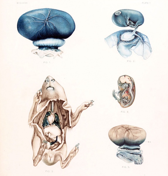
Plate 1
Fig. 1. Gross appearance of the placenta and fetal membranes from the uterus, 12 hours after injection of a vital stain into the amniotic cavity. Note the prominent omphalo-mesenteric vessels.
Fig. 2. Gross appearance of a guinea-pig fetus with the amnion opened 36 hours after injection of trypan-blue into the amniotic cavity. The omphalo-mesenteric vessels can be seen in the wall of the amniotic sac.
Fig. 3. Guinea-pig fetus, nearly full term, after injection of potassium ferrocyanide and iron ammonium citrate into the amniotic cavity. The fetus was killed 30 minutes after injection and immersed in acid formalin.
Notice the deep Prussian-blue stain in the lungs and trachea. The stomach contents also were blue. The kidneys are slightly stained.
Fig. 4. Sagittal section of guinea-pig, measuring 36 mm., 48 hours after the introduction of trypan-blue into the amniotic cavity. Note that the central nervous system remains unstained.
Fig. 5. Appearance of the placenta and membranes of a guinea-pig fetus after a single injection of trypan-blue into the maternal circulation. Notice the endodermal villi which are deeply stained, e. v. endodermal villi
| Historic Disclaimer - information about historic embryology pages |
|---|
| Pages where the terms "Historic" (textbooks, papers, people, recommendations) appear on this site, and sections within pages where this disclaimer appears, indicate that the content and scientific understanding are specific to the time of publication. This means that while some scientific descriptions are still accurate, the terminology and interpretation of the developmental mechanisms reflect the understanding at the time of original publication and those of the preceding periods, these terms, interpretations and recommendations may not reflect our current scientific understanding. (More? Embryology History | Historic Embryology Papers) |
File history
Click on a date/time to view the file as it appeared at that time.
| Date/Time | Thumbnail | Dimensions | User | Comment | |
|---|---|---|---|---|---|
| current | 17:45, 27 December 2012 |  | 1,145 × 1,200 (173 KB) | Z8600021 (talk | contribs) |
You cannot overwrite this file.
File usage
The following page uses this file:
