File:Windle1940 fig59-61.jpg
From Embryology

Size of this preview: 410 × 599 pixels. Other resolution: 1,391 × 2,032 pixels.
Original file (1,391 × 2,032 pixels, file size: 275 KB, MIME type: image/jpeg)
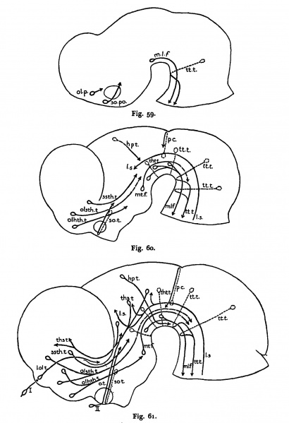
Figs. 59-61. Diagrams of the brains of cat embryos
Cat embryos 7 mm. (Fig. 59) , 10 mm. (Fig. 60) and 15 mm. (Fig. 61) C. R. length showing the principal über tracts present in each. Crossing neurons are dotted lines. Questionable courses: dash lines.
| Name of fibre group | Abbreviations used in figs. 59-61 | Size of smallest embryo (mm) in which it was found |
|---|---|---|
| Medial longitudinal fascicle | m.l.f. | 5.0 |
| Supraoptic system: direct preoptic component | so. po. | 6.0 |
| Supraoptic system: commissural component | so. t. | 7.0 |
| Olfacto-hypothalamic fibers | olhth. t. | 7.0 |
| Olfacto-subthalamic fibers | olsth. t. | 8.0 |
| Strio-subthalamic fibers | ssth. t. | 8.0 |
| Direct subthalamo-tegmental fibers (diffuse) | 8.0 | |
| Crossed pretecto-tegmental and thalamo-tegmental fibers (ventral commissure) | tho t. j tht. t. | 8.0 |
| Lemniscus system | l. s. | 8.0 |
| Terminal nerve fibers | 10.0 | |
| Posterior commissure fibers | p. c. | 10.0 |
| Habenulo-peduncular fibers | hp. t. | 10.0 |
| Lateral olfactory tract fibers | l. ol. t. | 11.5 |
| Mammillo-tegmental tract fibers | mt. f. | 11.5 |
| Optic nerve fibers | II | 11.5 |
| Thalamo-strial and thalamo-cortical fibers | ths. t. | 11.5 |
| Mammillo-thalamic fibers | 13.5 | |
| Olfactory nerve fibers | I | 13.0 |
Reference
Windle WF. Physiology of the Fetus. (1940) Saunders, Philadelphia.
Cite this page: Hill, M.A. (2024, April 25) Embryology Windle1940 fig59-61.jpg. Retrieved from https://embryology.med.unsw.edu.au/embryology/index.php/File:Windle1940_fig59-61.jpg
- © Dr Mark Hill 2024, UNSW Embryology ISBN: 978 0 7334 2609 4 - UNSW CRICOS Provider Code No. 00098G
File history
Click on a date/time to view the file as it appeared at that time.
| Date/Time | Thumbnail | Dimensions | User | Comment | |
|---|---|---|---|---|---|
| current | 10:07, 10 September 2018 |  | 1,391 × 2,032 (275 KB) | Z8600021 (talk | contribs) | |
| 10:06, 10 September 2018 |  | 1,391 × 2,274 (349 KB) | Z8600021 (talk | contribs) | ==Figs. 59-61. Diagrams of the brains of cat embryos== Cat embryos 7 mm. (Fig. 59) , 10 mm. (Fig. 60) and 15 mm. (Fig. 61) C. R. length showing the principal über tracts present in each. Crossing neurons are dotted lines. Questionable courses: dash l... |
You cannot overwrite this file.
File usage
The following page uses this file: