File:Wen1928-Fig06.jpg

Original file (1,280 × 917 pixels, file size: 280 KB, MIME type: image/jpeg)
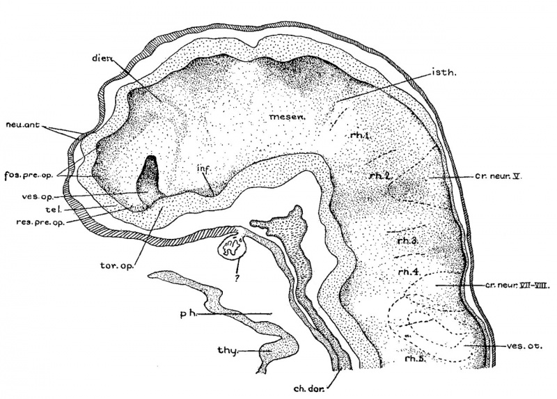
Fig. 6 A midsagittal projection reconstruction of the 22-somite embryo
H1093, showing most of the head. ‘ ?’ indicates the probable remains of the oral membrane.
In the 22-somite embryo, as in Davis’ 20-somite specimen, the closure of the anterior neuropore is completed, but its site on the external surface can still be recognized. The head ectoderm at that region is attached to the underlying brain wall in a small area probably representing the last point where the tissues separate (fig. 6).
| Abbreviations for all Figures | |
|---|---|
|
|
| Historic Disclaimer - information about historic embryology pages |
|---|
 |
- Links: fig 1 | Plate 2 | fig 2 | fig 3 | fig 4 | fig 5 | fig 6 | fig 7 | fig 8 | fig 9 | fig 10 | fig 11 | fig 12 | fig 13 | fig 14 | fig 15 | fig 16 | fig 17 | fig 18 | fig 19 | fig 21 | fig 21 | fig 22 | fig 23 | fig 24 | fig 25 | fig 26 | fig 27 | fig 28 | fig 29 | Wen 1928 | Carnegie stage 11 | Carnegie stage 12 | Historic Papers
Reference
Wen IC. The anatomy of human embryos with seventeen to twenty-three pairs of somites (1928) J. Comp. Neural., 45: 301-376.
Cite this page: Hill, M.A. (2024, April 23) Embryology Wen1928-Fig06.jpg. Retrieved from https://embryology.med.unsw.edu.au/embryology/index.php/File:Wen1928-Fig06.jpg
- © Dr Mark Hill 2024, UNSW Embryology ISBN: 978 0 7334 2609 4 - UNSW CRICOS Provider Code No. 00098G
File history
Click on a date/time to view the file as it appeared at that time.
| Date/Time | Thumbnail | Dimensions | User | Comment | |
|---|---|---|---|---|---|
| current | 14:26, 20 April 2016 | | 1,280 × 917 (280 KB) | Z8600021 (talk | contribs) | |
| 14:16, 20 April 2016 | 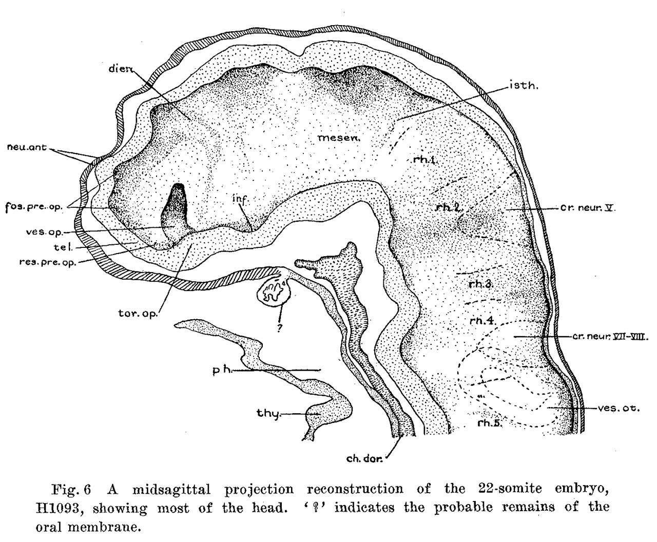 | 1,280 × 1,056 (311 KB) | Z8600021 (talk | contribs) |
You cannot overwrite this file.
File usage
The following page uses this file: