File:Wen1928-Fig05.jpg
From Embryology
Size of this preview: 512 × 599 pixels. Other resolution: 1,200 × 1,404 pixels.
Original file (1,200 × 1,404 pixels, file size: 288 KB, MIME type: image/jpeg)
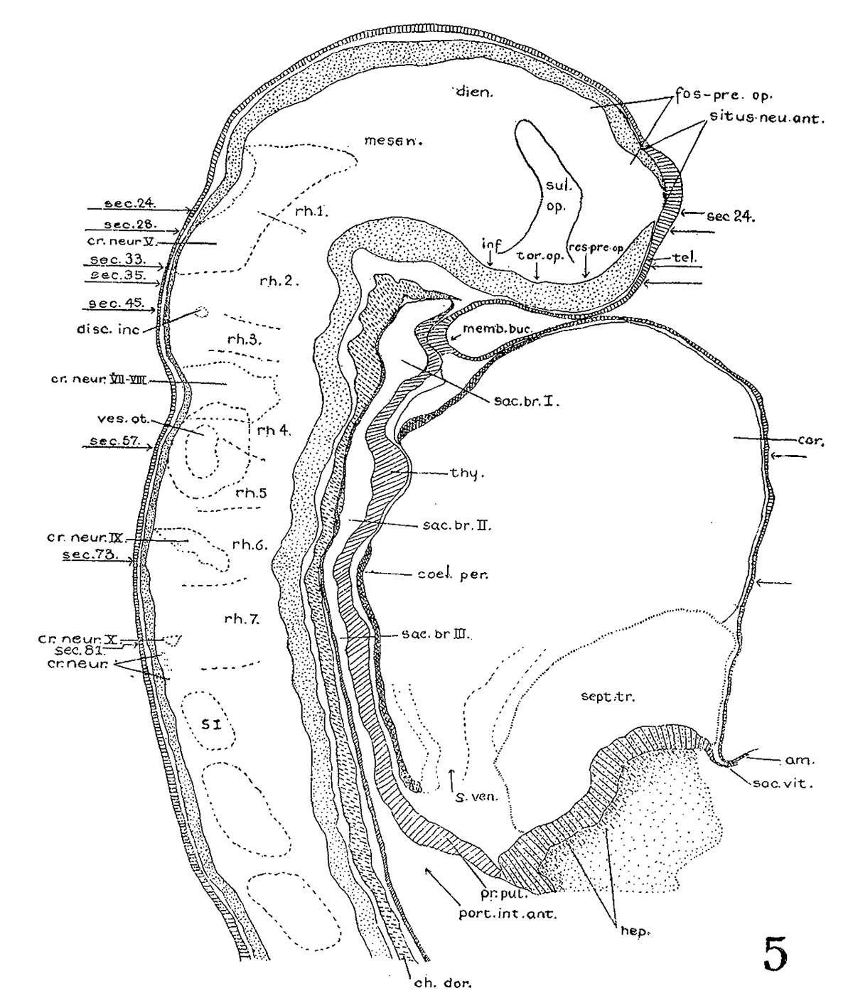
Fig. 5. Key drawing to Head of the 17-somite embryo H951
Key drawing to figure 4.
Prepared from a graphic reconstruction projected on the mid-sagittal plane.
| Abbreviations for all Figures | |
|---|---|
|
|
| Historic Disclaimer - information about historic embryology pages |
|---|
 |
- Links: fig 1 | Plate 2 | fig 2 | fig 3 | fig 4 | fig 5 | fig 6 | fig 7 | fig 8 | fig 9 | fig 10 | fig 11 | fig 12 | fig 13 | fig 14 | fig 15 | fig 16 | fig 17 | fig 18 | fig 19 | fig 21 | fig 21 | fig 22 | fig 23 | fig 24 | fig 25 | fig 26 | fig 27 | fig 28 | fig 29 | Wen 1928 | Carnegie stage 11 | Carnegie stage 12 | Historic Papers
Reference
Wen IC. The anatomy of human embryos with seventeen to twenty-three pairs of somites (1928) J. Comp. Neural., 45: 301-376.
Cite this page: Hill, M.A. (2024, April 18) Embryology Wen1928-Fig05.jpg. Retrieved from https://embryology.med.unsw.edu.au/embryology/index.php/File:Wen1928-Fig05.jpg
- © Dr Mark Hill 2024, UNSW Embryology ISBN: 978 0 7334 2609 4 - UNSW CRICOS Provider Code No. 00098G
File history
Click on a date/time to view the file as it appeared at that time.
| Date/Time | Thumbnail | Dimensions | User | Comment | |
|---|---|---|---|---|---|
| current | 15:06, 20 April 2016 | | 1,200 × 1,404 (288 KB) | Z8600021 (talk | contribs) | |
| 15:06, 20 April 2016 | 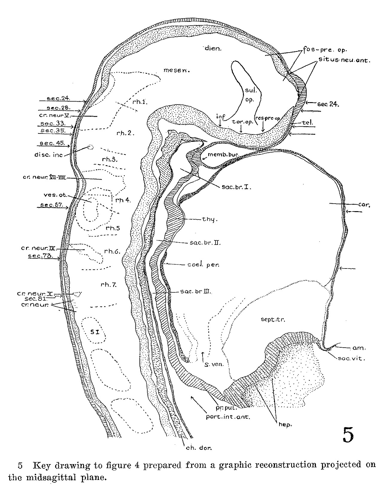 | 1,280 × 1,644 (366 KB) | Z8600021 (talk | contribs) |
You cannot overwrite this file.
File usage
The following page uses this file: