File:Wen1928-Fig03.jpg
Original file (809 × 900 pixels, file size: 116 KB, MIME type: image/jpeg)
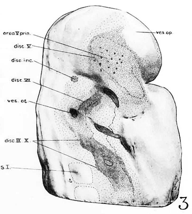
Fig 3 Photographs of the right side of a model of the head of the 17 somite embryo
Reduced in reproduction to 66% diameters. The ectodermal areas were projected from the model on which they had been plotted from measurements on the photomicrographs of the sections. The thickness of the ectoderm is indicated by the stippling, which is closer in the thicker parts. The coarse dots in area 17 pram. mark the location of cells emigrating from the placode; the spot marked * is illustrated in figure 11. The broken lines mark the areas over which deeply staining laminae of cells, probably of neural-crest origin, are closely applied to the overlying ectoderm. The visceral membranes are marked by parallel lines; sac.br.III indicates the position of the third pharyngeal pouch, which has not yet reached the ectoderm.
| Abbreviations for all Figures | |
|---|---|
|
|
| Historic Disclaimer - information about historic embryology pages |
|---|
 |
- Links: fig 1 | Plate 2 | fig 2 | fig 3 | fig 4 | fig 5 | fig 6 | fig 7 | fig 8 | fig 9 | fig 10 | fig 11 | fig 12 | fig 13 | fig 14 | fig 15 | fig 16 | fig 17 | fig 18 | fig 19 | fig 21 | fig 21 | fig 22 | fig 23 | fig 24 | fig 25 | fig 26 | fig 27 | fig 28 | fig 29 | Wen 1928 | Carnegie stage 11 | Carnegie stage 12 | Historic Papers
Reference
Wen IC. The anatomy of human embryos with seventeen to twenty-three pairs of somites (1928) J. Comp. Neural., 45: 301-376.
Cite this page: Hill, M.A. (2024, April 24) Embryology Wen1928-Fig03.jpg. Retrieved from https://embryology.med.unsw.edu.au/embryology/index.php/File:Wen1928-Fig03.jpg
- © Dr Mark Hill 2024, UNSW Embryology ISBN: 978 0 7334 2609 4 - UNSW CRICOS Provider Code No. 00098G
File history
Click on a date/time to view the file as it appeared at that time.
| Date/Time | Thumbnail | Dimensions | User | Comment | |
|---|---|---|---|---|---|
| current | 14:45, 20 April 2016 | | 809 × 900 (116 KB) | Z8600021 (talk | contribs) |
You cannot overwrite this file.
File usage
The following page uses this file: