File:WellsKaiser1959 fig02.jpg
From Embryology

Size of this preview: 512 × 599 pixels. Other resolution: 1,280 × 1,498 pixels.
Original file (1,280 × 1,498 pixels, file size: 399 KB, MIME type: image/jpeg)
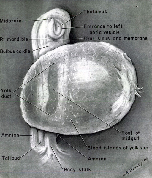
Fig. 2. Case 1. Ventrolateral aspect of the embryo
Ventrolateral aspect of the embryo, together with the amnion, yolk sac, and body stalk, when transilluminatecl with horizontal beams of light and viewed in the field of a dissecting microscope.
Reference
Wells LJ. and Kaiser IH Two Choice Human Embryos at Streeter’s Horizons XI and XIV. (1959)
Cite this page: Hill, M.A. (2024, April 25) Embryology WellsKaiser1959 fig02.jpg. Retrieved from https://embryology.med.unsw.edu.au/embryology/index.php/File:WellsKaiser1959_fig02.jpg
- © Dr Mark Hill 2024, UNSW Embryology ISBN: 978 0 7334 2609 4 - UNSW CRICOS Provider Code No. 00098G
File history
Click on a date/time to view the file as it appeared at that time.
| Date/Time | Thumbnail | Dimensions | User | Comment | |
|---|---|---|---|---|---|
| current | 11:59, 19 August 2017 |  | 1,280 × 1,498 (399 KB) | Z8600021 (talk | contribs) | |
| 11:58, 19 August 2017 |  | 1,749 × 2,087 (918 KB) | Z8600021 (talk | contribs) |
You cannot overwrite this file.
File usage
The following page uses this file: