File:Venule microvessel EM.jpg

Original file (600 × 626 pixels, file size: 91 KB, MIME type: image/jpeg)
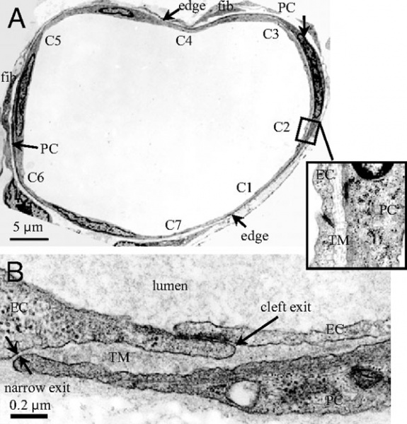
Rat Venular Microvessel Electron Micrograph
A Cross-section of a venular microvessel (VM) in rat mesentery. There are seven endothelial cell (EC) clefts, C1-C7, with all of the cleft exits covered by neighboring pericytes (PCs). PCs are immediately underneath ECs. There are two fibroblasts (fib.) outside PCs. The blowout shows C2 covered by a PC with a trapped microdomain (TM) of uniform thickness in between.
B An enlarged view showing a cleft exit covered by a PC, creating a TM of roughly uniform thickness except at the narrow exit.
Reference
<pubmed>18216252</pubmed>| PNAS
Copyright
Proceedings National Academy of Sciences (PNAS) Liberalization of PNAS copyright policy: Noncommercial use freely allowed Note original Author should be contacted for permission to reuse for Educational purposes. See also PNAS Author Rights and Permission FAQs
- Cozzarelli NR, Fulton KR, Sullenberger DM. Liberalization of PNAS copyright policy: noncommercial use freely allowed. Proc Natl Acad Sci U S A. 2004 Aug 24;101(34):12399. PMID15314225 "Our guiding principle is that, while PNAS retains copyright, anyone can make noncommercial use of work in PNAS without asking our permission, provided that the original source is cited."
Fig. 2.
File history
Click on a date/time to view the file as it appeared at that time.
| Date/Time | Thumbnail | Dimensions | User | Comment | |
|---|---|---|---|---|---|
| current | 13:40, 12 February 2012 | | 600 × 626 (91 KB) | S8600021 (talk | contribs) | ==Electron microscope images of a rat venular microvessel== (A) Cross-section of a venular microvessel (VM) in rat mesentery. There are seven endothelial cell (EC) clefts, C1-C7, with all of the cleft exits covered by neighboring pericytes (PCs). PCs are |
You cannot overwrite this file.
File usage
There are no pages that use this file.