File:Vagina histology 02.jpg
From Embryology
Size of this preview: 750 × 600 pixels. Other resolution: 1,280 × 1,024 pixels.
Original file (1,280 × 1,024 pixels, file size: 555 KB, MIME type: image/jpeg)
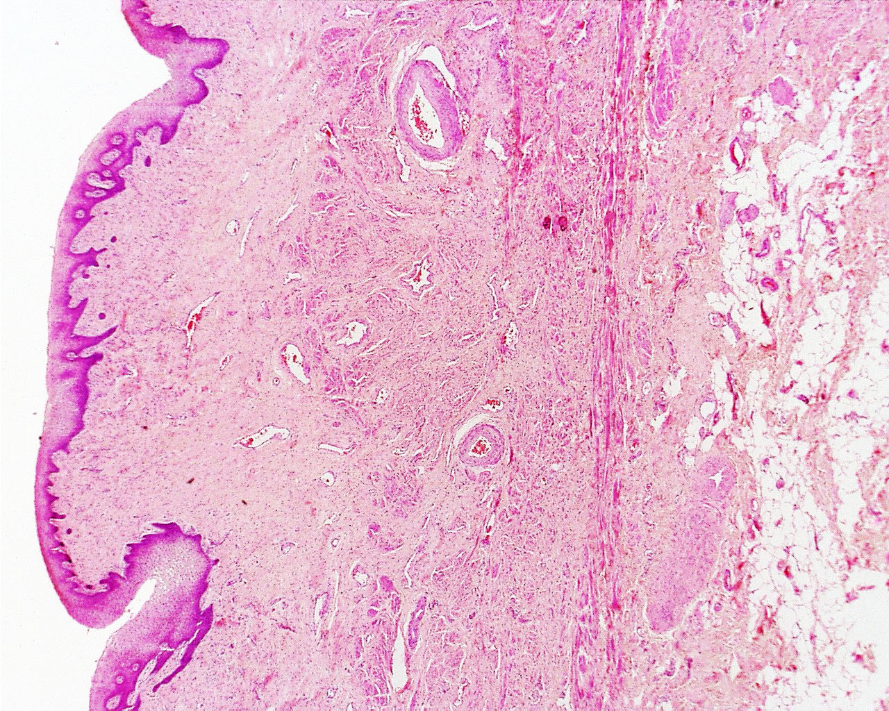
Vagina Histology Overview
(Stain - Haematoxylin Eosin)
The wall of the vagina fibromuscular tube consists of 3 layers: mucosa, muscularis and adventitia.
Mucosa
- stratified squamous epithelium (deep stratum basalis, intermediate stratum spinosum, superficial layers of flat eosinophilic cells
- rests on a very cellular lamina propria (many leukocytes).
- towards the muscularis some vascular cavernous spaces may be seen (erectile tissue).
Muscularis
- inner circular and outer longitudinal layers of smooth muscle
- bulbospongiosus muscle - (striated) forms an inferior sphincter around the vagina
Adventitia
- adventitia bordering the muscularis is fairly dense and contains many elastic fibres.
- outer part of the adventitia loose connective tissue with a prominent venous plexus.
File history
Click on a date/time to view the file as it appeared at that time.
| Date/Time | Thumbnail | Dimensions | User | Comment | |
|---|---|---|---|---|---|
| current | 09:51, 8 May 2013 | | 1,280 × 1,024 (555 KB) | Z8600021 (talk | contribs) | ==Vagina Histology== |
You cannot overwrite this file.
File usage
There are no pages that use this file.