File:Ultrasound - Hypoplastic left heart syndrome 02.jpg
Original file (890 × 626 pixels, file size: 54 KB, MIME type: image/jpeg)
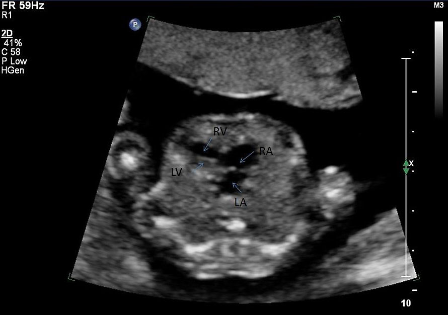
Ultrasound Hypoplastic Left Heart Syndrome
GA 19 week (second trimester) = week 17
The four chamber heart view shows a small left atrium (LA) and tiny left ventricle (LV).
LA - left atria
RA - right atria
LV - left ventricle
RV - right ventricle
- Links: Hypoplastic Left Heart | Ultrasound
Dr Stanley Ng - Obstetrical and gynecological sonologist (Sydney) for providing fetal ultrasound images and movie clips.
Cite this page: Hill, M.A. (2024, April 19) Embryology Ultrasound - Hypoplastic left heart syndrome 02.jpg. Retrieved from https://embryology.med.unsw.edu.au/embryology/index.php/File:Ultrasound_-_Hypoplastic_left_heart_syndrome_02.jpg
- © Dr Mark Hill 2024, UNSW Embryology ISBN: 978 0 7334 2609 4 - UNSW CRICOS Provider Code No. 00098G
File history
Click on a date/time to view the file as it appeared at that time.
| Date/Time | Thumbnail | Dimensions | User | Comment | |
|---|---|---|---|---|---|
| current | 16:18, 22 June 2016 | | 890 × 626 (54 KB) | Z8600021 (talk | contribs) | ==Ultrasound Hypoplastic Left Heart Syndrome== {{GA}} 19 week (second trimester) = week 17 LA - left atria RA - right atria LV - left ventricle RV - right ventricle :'''Links:''' [[Cardiovascular System - Hypoplastic Left Heart|Hypoplastic Left... |
You cannot overwrite this file.
File usage
The following page uses this file: