File:Thyng1914 plate5a.jpg
From Embryology

Size of this preview: 645 × 600 pixels. Other resolution: 1,613 × 1,500 pixels.
Original file (1,613 × 1,500 pixels, file size: 257 KB, MIME type: image/jpeg)
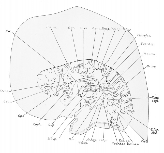
Plate 5. Reconstruction to show the lateral aspect of the left cephalic and cervical veins
Reconstruction to show the lateral aspect of the left cephalic and cervical veins, and the left jugular lymph sac of a 17.8 mm. human embryo (H. E. C. 839). X 11.9 diams.
- Embryo 17.8 mm Links: Fig 1 | Fig 2 | Plate 1a | Plate 1b | Plate 2a | Plate 2b | Plate 3a | Plate 3b | Plate 4a | Plate 4b | Plate 5a | Plate 5b | Plate 6 | Harvard Collection | Carnegie stage 19
Reference
Thyng FW. The anatomy of a 17.8 mm human embryo. (1914) Amer. J Anat. 17: 31-112.
Cite this page: Hill, M.A. (2024, April 24) Embryology Thyng1914 plate5a.jpg. Retrieved from https://embryology.med.unsw.edu.au/embryology/index.php/File:Thyng1914_plate5a.jpg
- © Dr Mark Hill 2024, UNSW Embryology ISBN: 978 0 7334 2609 4 - UNSW CRICOS Provider Code No. 00098G
File history
Click on a date/time to view the file as it appeared at that time.
| Date/Time | Thumbnail | Dimensions | User | Comment | |
|---|---|---|---|---|---|
| current | 19:21, 17 April 2014 |  | 1,613 × 1,500 (257 KB) | Z8600021 (talk | contribs) | ==Plate 5== {{Thyng1914 figures}} |
You cannot overwrite this file.
File usage
The following 3 pages use this file: