File:Thyng1914 plate4b.jpg
Original file (1,142 × 1,500 pixels, file size: 336 KB, MIME type: image/jpeg)
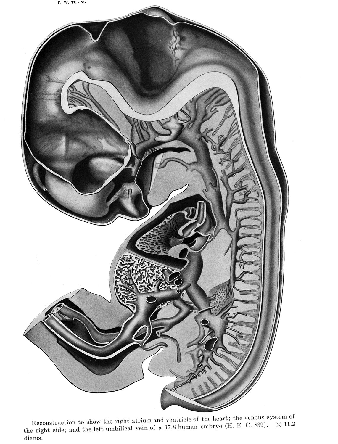
Plate 4. Heart, Right Venous System and Left Placental Vein
Reconstruction to show the right atrium and ventricle of the heart; the venous system of the right side; and the left umbilical vein of a 17.8 human embryo (H. E. C. 8.39). X 11.2 diams.
Neural
- The motor fibers issue from the metencephalon at a point slightly ventro-cephalad of the sensory root. They form a trunk of considerable size which crosses the medial surface of the semilunar ganglion to join the mandibular nerve (plate 4).
- The medial aspect of the right otic vesicle is represented in in plate 4.
Oral cavity
- The primitive oral cavity appears in median sagittal section in plate 1, but is represented more fully in plate 4, a portion of the tongue having been cut away.
- Process of the right maxillary arch is clearly shown in plate 4 where a portion of the tongue has been removed.
- Hypophysis (Hyp.), alluded to above, consists of a distal spade-like portion, connected to the oral epithelium in the median line by a slender stalk with reduced lumen. It is represented in side view in plate 4.
Heart
- In plate 4 the section passes through the cavities of the right side. The ventricular trabeculae are represented somewhat diagrammatically.
- Upon the dorsal wall of the right atrium (At.d., plate 4) is the sagittally directed, sinu-atrial orifice.
- The aortic septum {S.) is practically complete at this stage, and is seen in plate 4, separating the root of the aorta from the conus arteriosus (Con.art.) of the right ventricle.
Liver - The position of the quadrate lobe can be determined from plate 2. It is situated to the right of the vesica fellea (Ves.fel.), lying between this and the umbilical vein (V.um.s., plate 4).
Adrenal - The right suprarenal is shown in plate 4. It seems very probable to the author that each of these main groups of cells has been partially isolated from the suprarenal gland of its side by the development of the large dorso-ventral segment of the supra-ureteral venous channel (plate 4) which now intervenes between them.
Spleen - is clearly recognizable as a small, protuberance of the mesogastrium, containing dense m.esenchyma. The tissue directly ventral to this protuberance, is permeated by a vascular network, supplied by the splenic artery, and drained by the splenic vein (V.li., plate 4).
Arteries
- The superior mesenteric artery (A.mes.s.) takes origin from the aorta approximately on a level with the 12th thoracic pair of dorsal intersegmental arteries. It extends ventrally between the caudal ends of the suprarenal glands (plate 4) into the dorsal mesentery.
- The sciatic (inferior gluteal and A. comitans n. ischiadica) is at first dorso-lateral to the corresponding vein {V.isch., plate 4) with which it continues into the posterior limb-bud. The tap between it and the femoral has already occurred.
Veins
- The general distribution of the veins of the right side of the body is shown in plate 4.
- Anterior and common cardinal system. Between the dorsomedial surfaces of the cerebral hemispheres (plate 4) appears the paired anlage of the superior sagittal sinus (S. sag. sup.).
- The vertebral veins of which the right (V.vei'.d.) is reconstructed in plate 4, take the major share in the drainage of the cervical intersegmental veins.
- Only the right posterior cardinal derivatives have been reconstructed (plate 4) .
- The entire vena cava inferior, as shown in plate 4, is composed, therefore, of four parts.
- Portal system. The left umbilical vein {V.um.s.) is represented in plate 4. In the liver it communicates with the hepatic sinusoids, its blood passing chiefly through an especially large sinusoid, the ductus venosus (D.v.) which joins the left side of the common hepatic vein (vena cava inferioris) .
- Embryo 17.8 mm Links: Fig 1 | Fig 2 | Plate 1a | Plate 1b | Plate 2a | Plate 2b | Plate 3a | Plate 3b | Plate 4a | Plate 4b | Plate 5a | Plate 5b | Plate 6 | Harvard Collection | Carnegie stage 19
Reference
Thyng FW. The anatomy of a 17.8 mm human embryo. (1914) Amer. J Anat. 17: 31-112.
Cite this page: Hill, M.A. (2024, April 25) Embryology Thyng1914 plate4b.jpg. Retrieved from https://embryology.med.unsw.edu.au/embryology/index.php/File:Thyng1914_plate4b.jpg
- © Dr Mark Hill 2024, UNSW Embryology ISBN: 978 0 7334 2609 4 - UNSW CRICOS Provider Code No. 00098G
File history
Click on a date/time to view the file as it appeared at that time.
| Date/Time | Thumbnail | Dimensions | User | Comment | |
|---|---|---|---|---|---|
| current | 07:49, 10 April 2014 | | 1,142 × 1,500 (336 KB) | Z8600021 (talk | contribs) |
You cannot overwrite this file.
File usage
The following 4 pages use this file: