File:Thymus structure and function cartoon01.jpg

Original file (1,000 × 827 pixels, file size: 162 KB, MIME type: image/jpeg)
Thymus Structure and Function
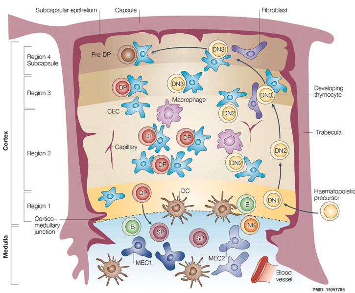
The thymus is broadly divided into two histologically defined regions, the cortex and the medulla, each of which contains several different thymic epithelial cell (TEC) subtypes.
In adults, T-cell precursors enter the thymus at the cortico–medullary junction, and then begin a highly ordered differentiation programme, which is linked to migration through the thymic stroma. Different thymocyte subsets are therefore found in spatially restricted regions of the thymus. The thymic cortex has been separated into four regions by Lind and colleagues8: region 1, the cortico–medullary junction, is the site of entry into the thymus and contains uncommitted progenitors, CD4-CD8- double-negative 1 (DN1) cells; in region 2, cells differentiate to the DN2 stage, undergo a proliferative clonal expansion, and lose B- and natural killer (NK)-cell potential; T-cell lineage commitment and the onset of T-cell receptor (TCR) beta-chain rearrangement occurs in DN3 cells in region 3; and the transition from DN to CD4+CD8+ double-positive (DP) status occurs in region 4. DP cells then migrate back through the cortex and, having differentiated into either CD4+ or CD8+ single-positive (SP) cells, into the medulla. Positive selection occurs mainly in the cortex, and requires cortical TECs, whereas negative selection occurs mainly in the medulla, and is mediated by medullary TECs and thymic dendritic cells (DCs). SP cells that have completed the differentiation programme egress from the medulla to the periphery. CEC, cortical epithelial cell; MEC, medullary epithelial cell. (text from figure 1 legend)
- Links: Thymus Development
Reference
<pubmed>15057786</pubmed>| Nat Rev Immunol.
Copyright
Reprinted by permission from Macmillan Publishers Ltd: [Nat Rev Immunol.] (Nature Reviews Immunology 4, 278-289 (April 2004) | doi:10.1038/nri1331), copyright (2004)
Cite this page: Hill, M.A. (2024, April 23) Embryology Thymus structure and function cartoon01.jpg. Retrieved from https://embryology.med.unsw.edu.au/embryology/index.php/File:Thymus_structure_and_function_cartoon01.jpg
- © Dr Mark Hill 2024, UNSW Embryology ISBN: 978 0 7334 2609 4 - UNSW CRICOS Provider Code No. 00098G
File history
Click on a date/time to view the file as it appeared at that time.
| Date/Time | Thumbnail | Dimensions | User | Comment | |
|---|---|---|---|---|---|
| current | 10:35, 25 February 2016 |  | 1,000 × 827 (162 KB) | Z8600021 (talk | contribs) | ==Thymus Structure and Function== The thymus is broadly divided into two histologically defined regions, the cortex and the medulla, each of which contains several different thymic epithelial cell (TEC) subtypes. In adults, T-cell precursors enter the... |
You cannot overwrite this file.
File usage
The following 2 pages use this file: