File:Thompson plate03.jpg
From Embryology
Size of this preview: 366 × 599 pixels. Other resolution: 679 × 1,112 pixels.
Original file (679 × 1,112 pixels, file size: 77 KB, MIME type: image/jpeg)
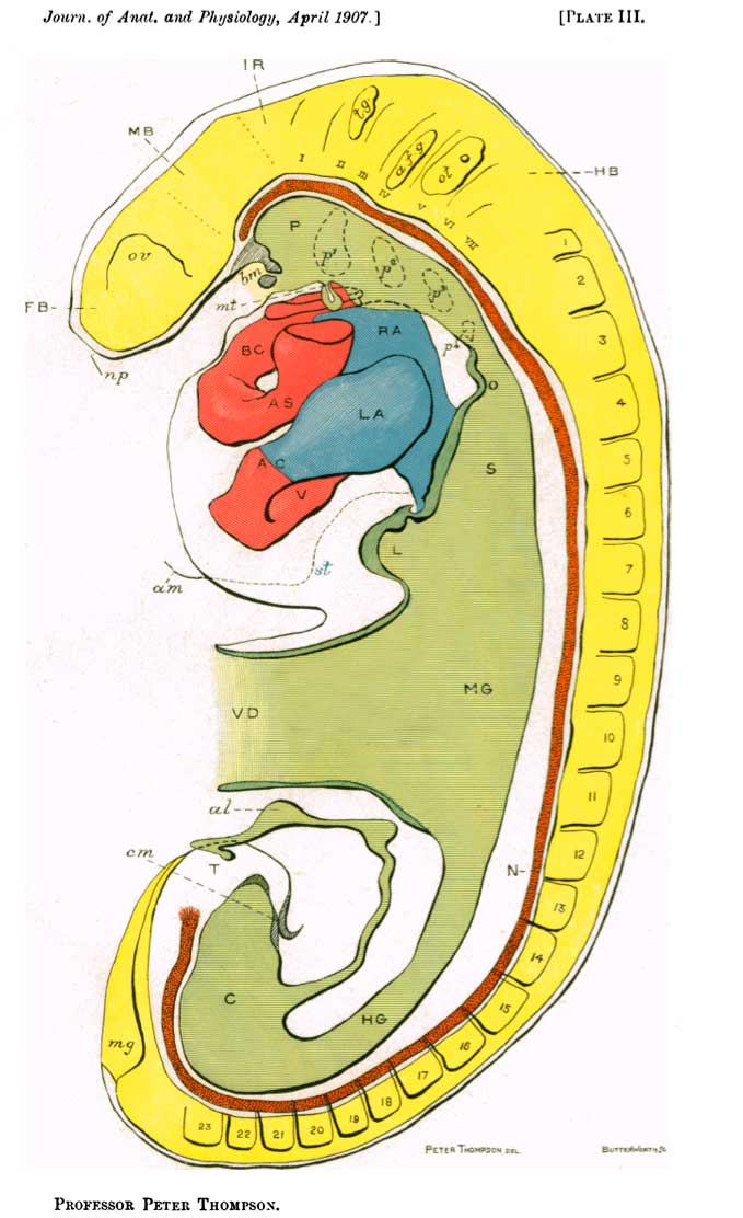
Plate III. Graphic reconstruction of embryo from serial sections
The endothelial tube only of the heart is shown.
Legend
- n.p., points to place of anterior neuropore
- F.B. - fore-brain
- o.v. - optic vesicle
- M.B. - mid-brain
- I.R. - isthmus rhombencephali
- i-vi - neuromeres of hind-brain H.B.
- t.g. - trigeminal ganglion
- a.f.g. - ganglion acoustico- facialis
- ot. - otocyst
- 1-23 somites
- b.m. - remains of bucco-pharyngeal membrane
- m.t. - median rudiment of thyroid body
- p.1-p.4 - pouches of pharynx P.
- o. - esophagus
- S. - stomach
- L. - liver
- s.t. - septum transversum
- am. - root of amnion
- V.D. - vitelline duct
- M.G. - mid gut
- N. - notoc1ord
- H.G. - hind gut
- C. - cloaca
- al. - allantois
- c.m. - cloacal membrane
- T. - tail
- m.g. - medullary groove
- R.A. and L.A. - right and left auricles
- v. - ventricle
- A.C. - auricular canal
- A.S. - aortic stem
- B.C. - bulbus cordis
(Note. - Neither the left horn of the sinus venosus nor the opening of the transverse piece into the right horn is indicated in the figure.)
- Links: Embryology History
| Historic Disclaimer - information about historic embryology pages |
|---|
 |
Reference
Thompson P. Description of a human embryo of twenty-three paired somites. (1907) J Anat Physiol, 41(3):159-71. PMID 17232726
File history
Click on a date/time to view the file as it appeared at that time.
| Date/Time | Thumbnail | Dimensions | User | Comment | |
|---|---|---|---|---|---|
| current | 15:41, 24 January 2012 | | 679 × 1,112 (77 KB) | S8600021 (talk | contribs) | :'''Links:''' Embryology History {{Historic Disclaimer}} ===Reference=== <pubmed>17232769</pubmed>| [http://www.ncbi.nlm.nih.gov/pmc/articles/PMC1289161 PMC1289161] Category:Historic Embryology |
You cannot overwrite this file.