File:ThompsonBrash1923 fig08.jpg
From Embryology
Size of this preview: 684 × 599 pixels. Other resolution: 1,395 × 1,222 pixels.
Original file (1,395 × 1,222 pixels, file size: 299 KB, MIME type: image/jpeg)
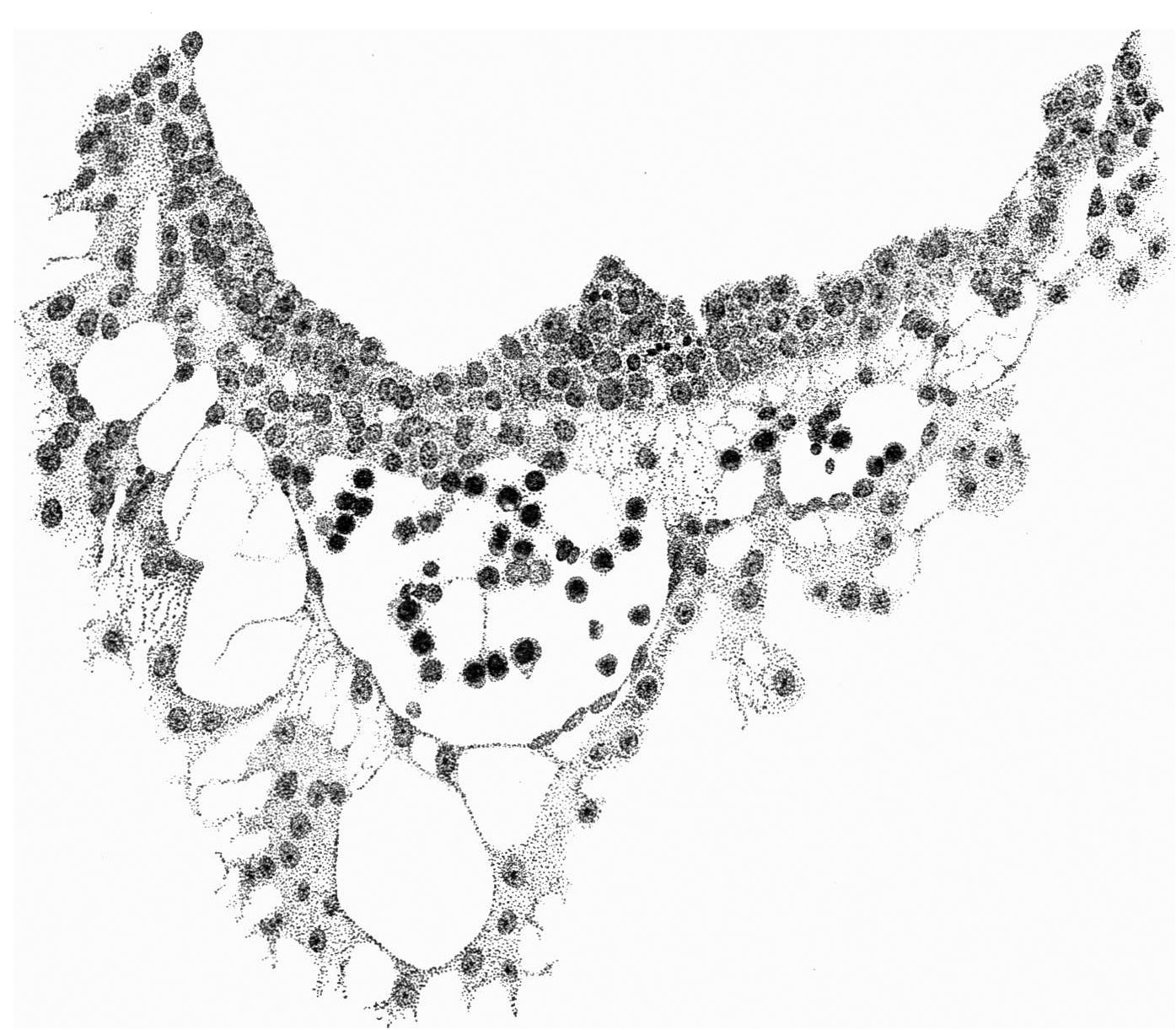
Fig. 8. The wall of the yolk-sac, near the cranial pole, showing blood-island
x 385.
| Historic Disclaimer - information about historic embryology pages |
|---|
 |
- Links: Fig. 1. | Fig. 2. | Fig. 3. | Fig. 4. | Fig. 5. | Fig. 6. | Fig. 7. | Fig. 8. | Fig. 9. | Fig. 10. | Fig. 11. | Fig. 12.
Reference
Thompson P. and Brash JC. A human embryo with head-process and commencing arch enteric canal. (1923) J Anat. 58: 1-20. PMID 17103992
Cite this page: Hill, M.A. (2024, April 24) Embryology ThompsonBrash1923 fig08.jpg. Retrieved from https://embryology.med.unsw.edu.au/embryology/index.php/File:ThompsonBrash1923_fig08.jpg
- © Dr Mark Hill 2024, UNSW Embryology ISBN: 978 0 7334 2609 4 - UNSW CRICOS Provider Code No. 00098G
File history
Click on a date/time to view the file as it appeared at that time.
| Date/Time | Thumbnail | Dimensions | User | Comment | |
|---|---|---|---|---|---|
| current | 17:55, 9 August 2015 | | 1,395 × 1,222 (299 KB) | Z8600021 (talk | contribs) | |
| 17:54, 9 August 2015 |  | 1,421 × 1,288 (280 KB) | Z8600021 (talk | contribs) | {{ThompsonBrash1923 figures}} |
You cannot overwrite this file.
File usage
The following page uses this file: