File:Thompson1908 fig01.jpg
Original file (1,134 × 865 pixels, file size: 295 KB, MIME type: image/jpeg)
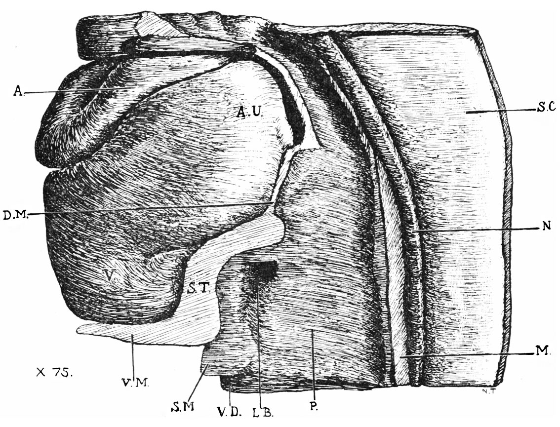
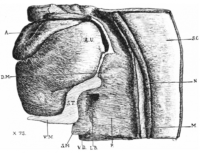
Fig 1. Part of model of embryo, with heart in situ
x 75.
S.C., spinal cord; N., notocord ; M., mesoderm between the two dorsal aortas ; A., aortic stem ; A.U., auricle ; D.M., dorsal mesocardium; S.T., septum trausversum; V.M,, ventral mesentery; S.M., wall of vitelline duct; V.D., cavity of vitelline duct lined by entoderm ; P., duodenum ; L.B., liver bud.
Fig. 1 is a drawing of a model reconstructed by the wax-plate method of a part of the embryo with the heart in situ. With the exception of the heart and spinal cord, the model is in sagittal section through the middle line, so that a view is obtained of the right half of the body from within. It shows the arrangement of a thick mesoblastic septum (S.T.) which is continuous cranialwards with the dorsal mesocardium and ventrally with the structure known as the ventral mesentery. The latter is continuous with the ventral body wall opposite the attachment of the root of the amnion. It will be clear, therefore, that the general disposition of the mesoderm here takes the form of an oblique septum, placed between the heart and the alimentary canal, and in front of the umbilical orifice. Leaving out of account the dorsal mesocardium, which subsequently disappears, the oblique septum consists of two parts: (1) a thick mass into which the liver bud is seen to be growing, and (2) the ventral mesentery. It is placed opposite the third and fourth cervical somites.
| Historic Disclaimer - information about historic embryology pages |
|---|
 |
Reference
Thompson P. A note on the development of the septum transversum and the liver. (1908) J Anat Physiol. 42(2): 170-5. PMID 17232762
Cite this page: Hill, M.A. (2024, April 18) Embryology Thompson1908 fig01.jpg. Retrieved from https://embryology.med.unsw.edu.au/embryology/index.php/File:Thompson1908_fig01.jpg
- © Dr Mark Hill 2024, UNSW Embryology ISBN: 978 0 7334 2609 4 - UNSW CRICOS Provider Code No. 00098G
File history
Click on a date/time to view the file as it appeared at that time.
| Date/Time | Thumbnail | Dimensions | User | Comment | |
|---|---|---|---|---|---|
| current | 22:49, 11 August 2015 | | 1,134 × 865 (295 KB) | Z8600021 (talk | contribs) | |
| 22:48, 11 August 2015 |  | 1,134 × 1,028 (335 KB) | Z8600021 (talk | contribs) |
You cannot overwrite this file.
File usage
The following page uses this file: