File:Thompson02.jpg
From Embryology

Size of this preview: 800 × 449 pixels. Other resolution: 1,049 × 589 pixels.
Original file (1,049 × 589 pixels, file size: 44 KB, MIME type: image/jpeg)
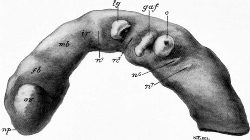
Fig. 2. Model of brain and part of the spinal cord
Original magnification x75.
Legend
- n.p. - place of anterior neuropore (closed)
- o.v. - optic vesicle
- f.b. - fore-brain
- m.b. - mid-brain
- i.r. - isthmus rhombeucephali
- n.1n.3,n,6 n.7 - neuromeres
- t.g. - trigenminal ganglion
- g.a.f. - ganglion acoustico-facialis
- o. - otocyst
- Links: Embryology History
| Historic Disclaimer - information about historic embryology pages |
|---|
 |
Reference
<pubmed>17232769</pubmed>| PMC1289161
File history
Click on a date/time to view the file as it appeared at that time.
| Date/Time | Thumbnail | Dimensions | User | Comment | |
|---|---|---|---|---|---|
| current | 15:39, 24 January 2012 | | 1,049 × 589 (44 KB) | S8600021 (talk | contribs) | :'''Links:''' Embryology History {{Historic Disclaimer}} ===Reference=== <pubmed>17232769</pubmed>| [http://www.ncbi.nlm.nih.gov/pmc/articles/PMC1289161 PMC1289161] Category:Historic Embryology |
You cannot overwrite this file.